An itchy speckled lesion
Case presentation
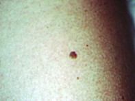
Over a two-month period, a 39-year-old woman noted the development of itching and darkening of an elevated pigmented lesion (Figure 1). The 0.5 cm lesion had appeared on her left arm only in the previous 12 months but had remained stable in size. Dermoscopy revealed a symmetrical lesion with extensive speckled pigment consisting of blue–black to yellow–white dots and globules. There was no evident pigment network and the central area was pink (Figure 2). The excision specimen showed verrucous epidermal hyperplasia with a prominent loose horny layer. There were several keratin-filled pseudocysts. The junctional zone of the hyperplastic epidermis was obscured by a very marked lymphocytic infiltrate associated with necrotic keratinocytes. Melanin pigment was present in the upper dermis (Figure 3).