Peer Reviewed
Perspectives on dermoscopy
Recurring pigmentation in an old scar
Abstract
Dermoscopy can be useful in evaluating recurrent pigmentation in surgical scars because it may highlight melanocytic proliferation.
Key Points
Case presentation
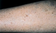
A 70-year-old woman had an 8 mm diameter mole removed from her left leg, and the mole was diagnosed histologically as a dysplastic junctional naevus. Eight years later, an irregular pigmented patch measuring 6 mm in diameter was noted at the lower border of the surgical scar (Figure 1). Dermoscopy revealed an asymmetrical, ill defined, broken pigment network with scattered small dark dots and isolated brown globules (Figure 2). Excision biopsy showed confluent proliferation of small hyperchromatic melanocytes in the junctional zone and focal collections of atypical melanocytes in the upper dermis (Figure 3).
Purchase the PDF version of this article
Already a subscriber? Login here.