Peer Reviewed
Perspectives on dermoscopy
A mole with a honeycomb pattern
Abstract
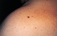
A patient presents with a flat pigmented mole on the back of the shoulder.
Key Points
Case presentation
A 48-year-old man had a longstanding mole on the back of his left shoulder (Figure 1). The mole measured 5 mm in diameter and had slowly increased in size. Dermoscopy revealed a dark brown reticulate network with a honeycomb pattern that faded at the periphery of the mole (Figure 2). Skin biopsy showed an epidermis with prominent elongated and narrow rete ridges. The rete ridges were deeply pigmented and contained increased numbers of melanocytes. In the underlying dermis there were numerous benign naevus cells which lacked pigment (Figure 3).
Purchase the PDF version of this article
Already a subscriber? Login here.