An irregular dark lesion of recent onset
Melanoma
Skin cancer
Case presentation
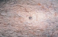
Over a six-month period, a 76-year-old man developed an irregular, deeply pigmented lesion (0.8 x 0.6 cm diameter) on the medial aspect of his right calf (Figure 1). Dermoscopy showed a pigmented lesion that was asymmetrical and had an ill-defined border. The pigment network was broad and highly irregular, associated with numerous dots and globules that ranged in colour from light brown to black. There was a patchy white veil and areas of hypopigmentation with blue–black dots (Figure 2). Excision biopsy showed an atrophic epidermis with confluent proliferation of atypical melanocytes that were present as single cells and nests along the epidermal junction (Figure 3). Within the upper dermis there were isolated clusters of atypical melanocytes as well as collections of macrophages packed with melanin (melanophages).