Peer Reviewed
Perspectives on dermoscopy
A symmetrical mole with asymmetrical pigment
Abstract
Clinically atypical moles may have no evident atypia on biopsy because the asymmetrical pigment of the mole can be created by diffuse pigment in the superficial dermal naevus cells.
Key Points
Case presentation
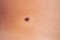
A 32-year-old woman with olive skin presented with an isolated, asymmetrically pigmented mole (measuring 0.6 by 0.4 cm) on her mid back (Figure 1). Dermoscopy revealed a symmetrically shaped mole that had a fine peripheral pigment network and a dark brown, partly homogeneous central zone with a jagged perimeter and patchy milky veil (Figure 2). Excision biopsy revealed an epidermis with an elongated pigmented rete ridge system containing isolated melanocyte nests (Figure 3). The dermis had a diffuse infiltrate of naevus cells. The superficial dermal naevus cells were pigmented.
Purchase the PDF version of this article
Already a subscriber? Login here.