Peer Reviewed
Perspectives on dermoscopy
A dark reticulated lesion masked by sun damage
Abstract
The diagnosis of pigmented lesions is a daily challenge in general practice. Dermoscopy can provide extra clues, but requires significant expertise. This series will help you hone your skills.
Key Points
Case presentation
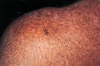
A 64-year-old man developed a 1.5 x 0.5 cm, irregularly shaped, pigmented lesion on his posterior left shoulder (Figure 1). He had mottled, chronic sun-damaged skin due to lifelong sun exposure. He was unaware of the presence of the lesion. Dermoscopy revealed an asymmetrical and irregularly bordered lesion with a light to dark brown reticular pigment network (Figure 2). The network was interrupted by multiple small flesh-coloured dots. A shave biopsy showed an epidermis with well developed and deeply pigmented rete ridges forming an antler-like pattern (Figure 3). There was no increase in number of melanocytes and no evident melanocytic nests or atypic.
Purchase the PDF version of this article
Already a subscriber? Login here.