Peer Reviewed
Perspectives on dermoscopy
Seborrhoeic keratosis with a black nodule
Abstract
Irritation of a seborrhoeic keratosis may produce a benign nodular black component that may be difficult to diagnose both clinically and with the dermoscope. Diagnosis may require skin biopsy.
Key Points
Case presentation
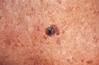
A 56-year-old man presented with a smooth shiny black nodule (1.8 cm in diameter) that was superimposed on a longstanding seborrhoeic keratosis on his chest wall (Figure 1). The nodule had appeared over a six-month period between skin checks. Dermoscopy revealed a central homogeneous jet black surface and irregular periphery that merged with a blue–grey honeycombed corona containing linear and small round sandy deposits. The outer margin had an irregular border with further dark centres that merged with mottled sun-damaged skin (Figure 2). Excision biopsy showed a seborrhoeic keratosis with a hyperplastic epidermis, hyperpigmentation, keratin pseudocysts and inflammation, but no atypia (Figure 3).
Purchase the PDF version of this article
Already a subscriber? Login here.