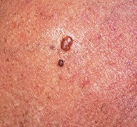
A pale papule with a pigmented ring
Melanoma
Skin cancer
Case presentation
A 48-year-old man noted the recent development of a 3 mm diameter pale papule with a pigmented ring over his left upper chest. Eighteen months previously, he had had a 5.6 mm thick amelanotic melanoma removed from the back of his scalp, and subsequently had deep wide excision of the site, followed by lymph node dissection and radiotherapy. Dermoscopy revealed a pale papule outlined by a rim of dark pigment with dark dots and a partial network. Skin biopsy showed a nodule composed of atypical melanocytes associated with peripheral intraepidermal extension. Melanin stain highlighted the peripheral hyperpigmentation seen clinically and under the dermatoscope. No other lesions were found on careful clinical review, but subsequently further metastases developed.