Peer Reviewed
Perspectives on dermoscopy
A recent pale nodule with pseudocysts
Abstract
The diagnosis of pigmented lesions is a daily challenge in general practice. Dermoscopy can provide extra clues, but requires significant expertise.
Key Points
Case presentation
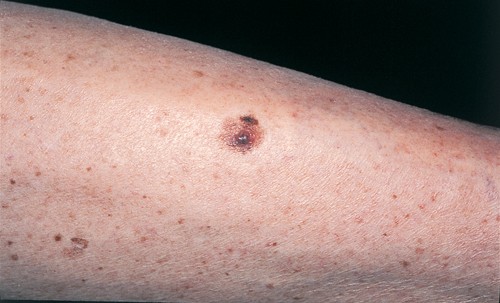
Over a one-month period, an 85-year-old man noted the development of a pale pigmented nodule in a tan-coloured macule that had been present for five years on his left shin. Dermoscopy revealed a pale nodule with isolated white pseudocysts and a partial blue–grey veil, and the nodule was surrounded by an asymmetrical pigment network with a broken irregular mesh. Biopsy of the nodule revealed sheets of atypical spindle cells with mitoses within the dermis and scant melanin pigment.
Purchase the PDF version of this article
Already a subscriber? Login here.