Peer Reviewed
Perspectives on dermoscopy
A hazy pigmented lesion
Abstract
What does dermoscopy reveal about this lesion?
Key Points
Case presentation
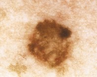
A 29-year-old woman had a longstanding 6 mm diameter irregularly pigmented mole on her upper back. Dermoscopy of the mole revealed hazy pigmentation with an asymmetrical pattern and a focally hyperpigmented focus at the upper right edge. There was an irregular pigment network with patchy small diameter mesh. The network at the periphery blended with the surrounding freckles. Excision biopsy of the lesion revealed an epidermis with a prominent elongated rete ridge system, which projected into the dermis as an anastomosing network. There were uniform pigmented melanocytes at the epidermal junction, and melanocytes were also present as clusters of small focally pigmented cells in the upper dermis.
Purchase the PDF version of this article
Already a subscriber? Login here.