Peer Reviewed
Perspectives on dermoscopy
A mole of recent onset
Recent articles on:
Moles
Moles
Abstract
This is another case to help you hone your skills in dermoscopy.
Key Points
Case history
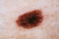
A 62-year-old man developed over his left lower abdomen a darkly pigmented symmetrical and oval mole that measured 5 x 3 mm (Figure 1). The mole was absent in baseline photographs taken two years before the consultation. Dermoscopy revealed a symmetrical lesion that had a well developed pigment network with a uniform pattern. The network was indistinct in the centre and had several pale ‘holes’ at the periphery (Figure 2). Excision biopsy showed an epidermis with a well formed pigmented rete ridge system associated with increased single melanocytes and nests of melanocytes that were not confluent. The upper dermis showed scattered lymphocytes and some melanin pigment (Figure 3).
Purchase the PDF version of this article
Already a subscriber? Login here.