Peer Reviewed
Perspectives on dermoscopy
A solitary pigmented nodule on the thigh
Abstract
This series will help clinicians with an interest in dermoscopy. This month, we present a 55-year-old woman with an asymptomatic nodule on her thigh.
Key Points
- With sufficient training and expertise, clinicians can use dermoscopy to improve diagnostic accuracy for melanocytic lesions and other common skin tumours.
Case presentation
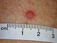
A 55-year-old woman presented with an asymptomatic pigmented nodule on her left thigh that had been present for six months (Figure 1). The lesion was approximately 6 mm in maximal diameter and firm on palpation. Lateral compression of the lesion resulted in dimpling of the nodule. Dermoscopy revealed a central, white, scar-like patch with a delicate peripheral pigment network in association with vertically orientated telangiectatic vessels (Figure 2).
Purchase the PDF version of this article
Already a subscriber? Login here.