A smudge on the sole – safe or sinister?
Foot injuries and disorders
Case presentation
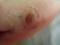
A 57-year-old Caucasian woman presented for a routine skin check. She had a pigmented lesion on the sole of her left foot, which she had circled so she would not forget to enquire about it. There was a past history of lentigo maligna and the patient, now aware that her risk of a second primary melanoma was increased, was anxious about each check up. She felt that the lesion had been stable and longstanding, but her husband insisted that it had grown over the years.
The lesion was a somewhat smudgy, light tan macule measuring 5 x 3 mm and was located just proximal to the base of the fifth toe on plantar skin (Figure 1a). There appeared to be some accentuation of pigment in a linear configuration. On dermoscopy the pigment was aligned in a lattice pattern and plantar eccrine gland openings were not observed (Figure 1b).