Managing patients after implantation of a cardiac rhythm management device

Pacemakers and Implantable Defibrillators
Arrhythmia
Patients with cardiac rhythm management devices such as pacemakers and defibrillators are cared for jointly by GPs and cardiologists. GPs can manage issues such as wound care, bleeding and pain, and provide guidance regarding work, travel and household appliance use.
- Implanted cardiac rhythm management (CRM) devices are commonly used in Australia.
- The main types of implanted devices are pacemakers, defibrillators, biventricular pacemakers/defibrillators and implantable loop recorders.
- Care of patients with CRM devices should be jointly shared between the GP and cardiologist.
- Early complications are best referred back to the interventional cardiologist; they include wound-related issues (active bleeding, infection, large haematoma), unusual postprocedural pain (pericardial or pleuritic pain) and noncardiac muscle twitching.
- There are many misconceptions in patients with implanted CRM devices regarding legal and safe driving, the use of household devices such as mobile phones and microwave ovens, foreign travel and working safely in environments where there are sources of electromagnetic interference.
Picture credit: © Aberration Films Ltd/SPL/Diomedia.com
Approximately 20,000 cardiac rhythm management (CRM) devices are implanted per year in patients in Australia.1 Although patients with these devices require lifelong cardiologist follow up, GPs are likely to encounter patients who are concerned about their devices, both immediately after implantation and later. This article discusses the common devices implanted and associated postprocedural issues, and also lifestyle guidance regarding work, travel and household appliance use for patients with these devices.
Device types and their implantation
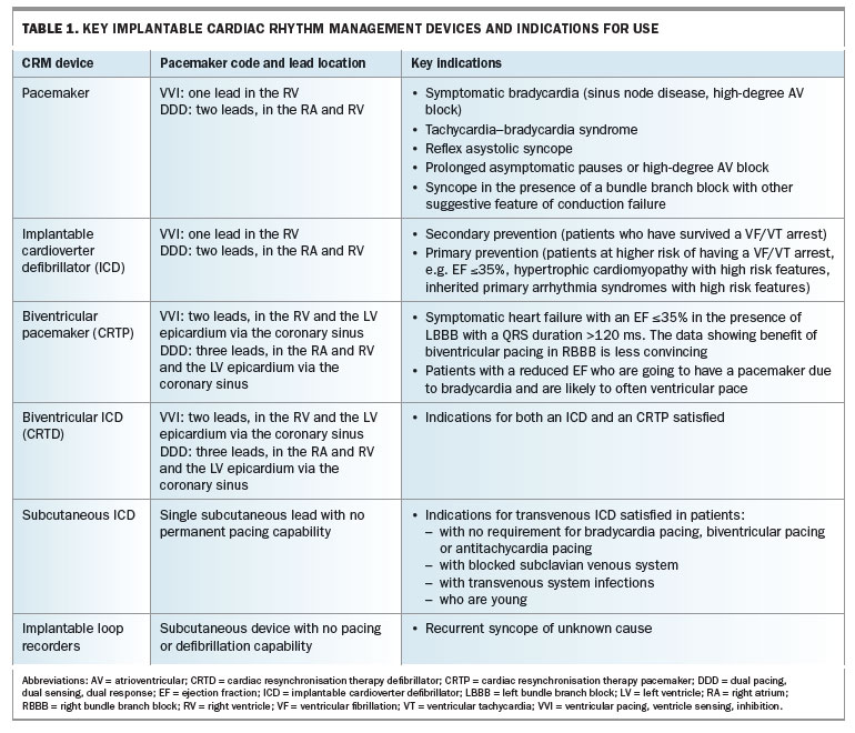
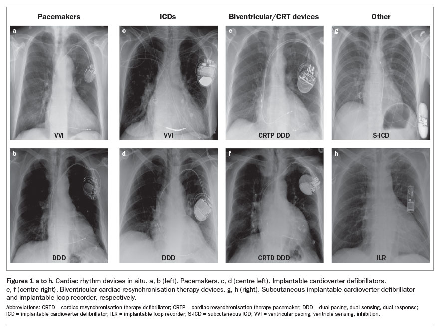
Comprehensive details regarding the indications of CRM devices are provided in international guidelines and are summarised in Table 1.2-4 Chest radiographs of patients with these devices in situ are shown in Figures 1a to h.
Pacemakers and defibrillators are composed of a battery-powered pulse generator attached to one, two or three leads inserted into the heart. The leads may be positioned in the right atrium or the right ventricle (single-chamber pacemaker) or both (dual-chamber pacemaker), and may also be attached to the left ventricular epicardium via the coronary sinus (to give biventricular pacing). A three-letter code defines the pacing mode of the pacemaker:
- the first letter denotes the chamber paced – Atrium, Ventricle or Dual
- the second letter, the chamber sensed – Atrium, Ventricle or Dual
- the third letter, how a pacemaker responds to a sensed event – I = sensed event Inhibits pacing, T = pacing Triggered in response to a sensed event, D = Dual modes of response.
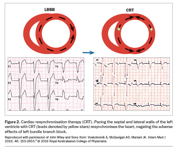
Cardiac resynchronisation therapy (CRT), also known as biventricular pacing, aims to reverse the deleterious effects of left bundle branch block in patients with heart failure and a reduced ejection fraction. The right and left ventricular pacing leads generate two ventricular activation wavefronts that move towards each other. The benefit of resynchronisation therapy lies in effective fusion of these two depolarisation wavefronts, synchronising the walls of the left ventricle, thereby improving efficiency of the heart’s pumping function (Figure 2).5 This benefit is greatest in patients with left bundle branch block.
Pacemakers, implantable cardioverter defibrillators (ICD), cardiac resynchronisation therapy pacemakers (CRTPs) and cardiac resynchronisation therapy defibrillators (CRTDs) are usually placed in the left infraclavicular region (occasionally on the right) and connected to the myocardium via transvenous leads. Most leads are actively attached to the myocardium (active fixation) – they have a small retractable helix (corkscrew) that allows anchoring. Passive fixation leads have tines to facilitate adherence to the myocardium. Some devices have no transvenous connection: subcutaneous defibrillators (S-ICDs) are placed in the left lateral chest wall with a subcutaneous tunnelled lead, and implantable loop recorders (ILR) are inserted in the left parasternal region and have no lead. Rarely, devices are implanted surgically with the leads directly attached to the epicardium, which has the advantage of not requiring a transvenous system but the disadvantage of a more invasive procedure under general anaesthetic. The epicardial technique is reserved for when there is difficulty in delivering a pacing lead to the intended cardiac chamber (e.g. in the presence of subclavian vein obstruction, congenital heart disease, challenging coronary sinus anatomy), cardiac surgery is being performed and there is a concurrent need for permanent pacing, there is a mechanical tricuspid valve or in certain paediatric populations.
Follow up after implantation
After a new device is implanted, patients are usually seen six weeks later and subsequent follow ups are every six to 12 months, guided by patient- and device- related factors.
Some patients also have remote monitoring, whereby the device wirelessly sends data relating to its function and recorded arrhythmias to the cardiologist. This monitoring requires a daily transmission of information by a landline or mobile telephone; mobile telephonic transmission of information may have limited applicability in rural areas because of poor mobile telephone reception. Remote monitoring allows rapid detection of abnormal device function and arrhythmia events and its uptake is likely to increase with time.6 Any device reprogramming, however, requires attendance at the pacemaker clinic.
Devices can also be programed to deliver a regular but not continuous audible/vibration alert that would be noticeable to the patient if a potential hazardous error is detected. If this occurs, the patient should see their cardiologist urgently for further assessment and interrogation of the device.
Post-implantation issues
Wound care
The surgical incision from device implantation is usually closed with absorbable stitches. However, if nonabsorbable sutures are used, they should be removed five to seven days after the procedure.
The patient should keep the incision dry, ideally with a waterproof dressing, for at least five days to minimise the risk of infection. During this period, options for bathing include showering with a detachable shower head directed away from the wound and a sponge bath, covering the dressing with a towel. If the wound becomes wet, the dressing should be removed and a hair dryer used to dry the skin before redressing, rather than rubbing with a towel.
For the first month, pressure on the wound should be minimised. Handbags, backpacks, brassiere straps, seat belts and slings should not traverse the incision.
Haematoma
The risk of pacemaker pocket haematoma is approximately 5% and its occurrence is dependent on both surgical technique and patient-related factors.7 A combination of antiplatelet agent use and preoperative heparin is associated with the highest risk.
Haematomas can be managed conservatively, provided there is no active bleeding and the skin sutures have integrity; very large haematomas can take up to six weeks to resorb. A patient with an actively bleeding wound that cannot be controlled by direct pressure should be referred back to the implanting centre for urgent review. Needle aspiration of the pocket should never be attempted as this may seed infection into a previously sterile pocket, with no diagnostic or therapeutic benefit for the patient. If there is any doubt, the patient should be referred back to the implanting centre.
Pain
Implant site pain is expected for the first few days after the implantation procedure and is best managed with paracetamol and intermittent application of cold compresses. Occasionally, stronger analgesia is required, particularly after a subpectoral muscle implant.
Significant pain occurring more than four weeks after implantation should prompt further evaluation to exclude cellulitis, pocket infection, impending erosion or superficial implant of the device. White blood cell counts and C-reactive protein levels may remain unremarkable even in the presence of a significant pocket infection. Therefore, prompt clinical evaluation in the pacemaker clinic is warranted. The incidence of infection is higher after a generator change (5.3 per 1000 patient-years) than after a new implant (1.8 per 1000 patient-years).8
Pleuritic or pericardial pain in the first month after the procedure requires immediate assessment to exclude pneumothorax (less than 1% risk) or lead perforation into the pericardium with or without tamponade (less than 0.5% risk).9 Deceivingly, these complications can occur despite initially reassuring predischarge pacing checks and chest radiographs. These complications are only seen when leads are placed in the heart, and are therefore not associated with generator replacements, implantable loop recorders or subcutaneous ICDs.
Muscle twitching
Electric impulses from CRM devices are expected to stimulate myocardium; however, they can also stimulate the diaphragm, intercostal or pectoral muscles. This muscle twitching may be benign (e.g. left ventricular lead in close proximity to the phrenic nerve) or indicate a problem such as lead displacement or insulation fracture. Patients with new or persistent noncardiac muscle stimulation should always be referred to the pacing clinic.
Deep venous thrombosis
Unilateral upper arm swelling associated with pain or erythema is suggestive of upper limb deep venous thrombosis, which can be confirmed by Doppler ultrasound or computed tomography venogram. The incidence of this complication in pacemaker implantation is less than 5%, although asymptomatic subclavian vein thrombosis is more common.10,11 Anticoagulation for at least three months is recommended if deep venous thrombosis is confirmed.
No improvement or recurrence in symptoms
Syncope in patients with an implanted pacemaker is not common but requires urgent action. It is reported as occurring in 18% of patients implanted for sinus node disease at 10 years of follow up and in 5% of those implanted for atrioventricular block at five years’ follow up.11 A pacemaker hardware fault (lead exit block, conductor fracture, insulation break, battery depletion) or software problem is the culprit in less than 5% of cases and is diagnosed by an urgent pacing check. The most common cause is vasovagal syncope, which is seldom alleviated by pacemaker programming.
In patients with ICDs, syncope, aborted syncope or sensation of defibrillation (commonly described by patients as ‘a horse kicking you in the chest’) may represent malignant ventricular arrhythmias and should be referred to the treating cardiologist and/or emergency department immediately.
Patients with sinus node dysfunction receiving a pacemaker may feel still nonspecifically flat after insertion of the device. Activation and optimisation of rate-responsive pacing is often necessary at the initial and subsequent visits.
The development of atrial fibrillation may not be obvious in the patient with a pacemaker because the usual finding of an irregular rhythm is masked by regular ventricular pacing; the clinical significance could be both stroke risk and unexplained symptoms of heart failure or effort intolerance. An ECG can be performed, and suspicion aroused if the ventricular rate is fixed on 70 beats per minute with no preceding ‘p’ wave, but often a visit to the pacemaker clinic is required.
Chronic right ventricular pacing can induce heart failure, known as pacing-induced cardiomyopathy, in up to 12% of patients over four years.12 If patients with pacemakers develop symptoms of heart failure and reduced ejection fraction is confirmed, a system upgrade with addition of a left ventricular lead can reverse the cardiomyopathy in up to 80% of cases.12
Cardiac resynchronisation therapy improves symptoms and mortality outcomes in selected patients with heart failure.5 Approximately 70% of patients experience an improvement with this therapy.5 Those who do not respond should have detailed cardiology assessment to optimise their resynchronisation therapy.
Lifestyle guidance
Driving
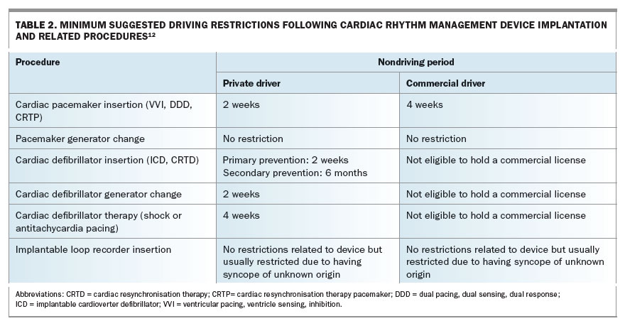
In Australia, it is mandatory that patients notify their state driving licensing authority following implantation of a CRM device.13 Patients are subject to driving restrictions after device implantation and other device-related procedures, with suggested minimum nondriving periods, and must remain free of syncope for this period before driving again (Table 2).13
Foreign travel
The manufacturers of CRM devices used in Australia have a worldwide presence, hence local healthcare providers will be able to help in the unlikely event of a device malfunction abroad. Nonetheless, a CRM device check before departure is recommended. The Implantable Device Identification Card contains information that may be invaluable at border security or in the event of a medical emergency abroad, and should be carried by the patient. These cards are mailed to the patient directly from the device manufacturer following an implant. Alternatively, the relevant data can be obtained from the pacemaker clinic.
Adequate travel insurance with full disclosure of medical conditions is likely to be the biggest limitation to foreign travel. Many insurers require 90 days of no change in medical condition, which can include medication dose changes, to issue complete cover.
Flying is safe if the patient is in status quo 48 hours after an uncomplicated CRM device implant. If the procedure was complicated by a pneumothorax, there should be a minimum period of two weeks for recovery.14
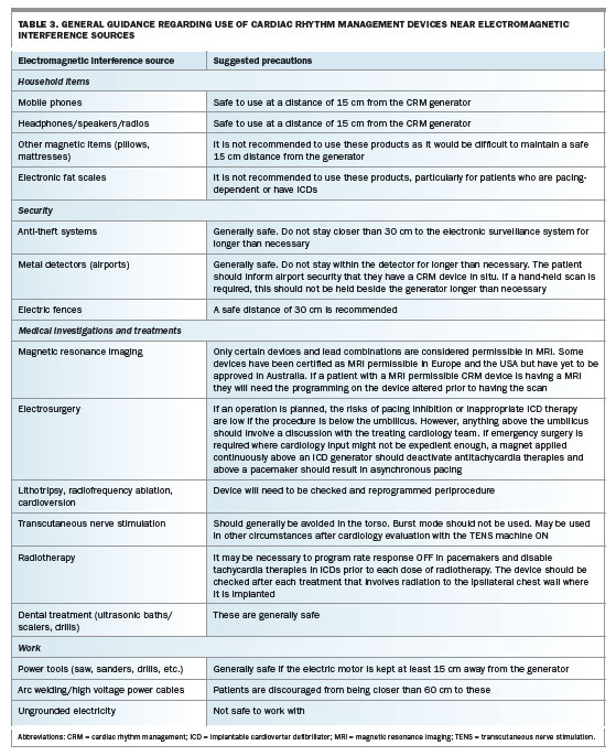
Electromagnetic interference
Tissue heating, circuit failure, inhibition of pacing output and ICD misfiring can result from electromagnetic interference affecting CRM devices.15 Modern device construction has mitigated the risk from most electromagnetic interference sources to minimal levels. General advice is given in Table 3 and also online (e.g. the American Heart Association website; search for ‘Devices that may interfere with pacemakers’ and ‘Devices that may interfere with implantable cardioverter defibrillators [ICDs]’).16-18 Potential risks are higher in patients with pacemakers who are pacing-dependent (have no underlying intrinsic heart rhythm) and in those with an ICD. There are no significant risks of electromagnetic interference with implantable loop recorders.
As the potential sources of electromagnetic interference increase (known and unknown) in both the workplace and home, unexpected interactions with CRM devices may occur. In certain situations it may be appropriate to enlist an approved contractor to perform environmental testing for electromagnetic interference that might affect CRM device function.
Conclusion
GPs and cardiologists care for patients with CRM devices jointly. GPs are likely to be consulted by patients with these devices regarding post-implantation issues such as wound care, bleeding and pain, and should feel empowered to provide lifestyle guidance to these patients with respect to work, travel and household appliance use. Complications or device malfunction can occur at any stage and, if suspected, should prompt referral to the patient’s cardiologist. MT
COMPETING INTERESTS: None.
References
1. Mond HG, Crozier I. The Australian and New Zealand cardiac pacemaker and implantable cardioverter-defibrillator survey: calendar year 2013. Heart Lung Circ 2015; 24: 291-297.
2. Brignole M, Auricchio A, Baron-Esquivias G, et al. 2013 ESC Guidelines on cardiac pacing and cardiac resynchronization therapy: the Task Force on cardiac pacing and resynchronization therapy of the European Society of Cardiology (ESC). Developed in collaboration with the European Heart Rhythm Association (EHRA). Eur Heart J 2013; 34: 2281-2329.
3. Priori SG, Blomstrom-Lundqvist C, Mazzanti A, et al. 2015 ESC Guidelines for the management of patients with ventricular arrhythmias and the prevention of sudden cardiac death: The Task Force for the Management of Patients with Ventricular Arrhythmias and the Prevention of Sudden Cardiac Death of the European Society of Cardiology (ESC). Eur Heart J 2015; 36: 2793-2867.
4. Epstein AE, DiMarco JP, Ellenbogen KA, et al. 2012 ACCF/AHA/HRS focused update incorporated into the ACCF/AHA/HRS 2008 guidelines for device-based therapy of cardiac rhythm abnormalities: a report of the American College of Cardiology Foundation/American Heart Association Task Force on Practice Guidelines and the Heart Rhythm Society. Circulation 2013; 127: e283-e352.
5. Voskoboinik A, McGavigan AD, Mariani JA. Cardiac resynchronisation therapy in 2015: keeping up with the pace. Intern Med J 2016; 46: 255-265.
6. Slotwiner D, Varma N, Akar JG, Annas G, Beardsall M, Fogel RI, et al. HRS Expert Consensus Statement on remote interrogation and monitoring for cardiovascular implantable electronic devices. Heart Rhythm 2015; 12:
e69-100.
7. Wiegand UK, LeJeune D, Boguschewski F, et al. Pocket hematoma after pacemaker or implantable cardioverter defibrillator surgery: influence of patient morbidity, operation strategy, and perioperative antiplatelet/anticoagulation therapy. Chest 2004; 126: 1177-1186.
8. Johansen JB, Jorgensen OD, Moller M, Arnsbo P, Mortensen PT, Nielsen JC. Infection after pacemaker implantation: infection rates and risk factors associated with infection in a population-based cohort study of 46299 consecutive patients. Eur Heart J 2011; 32: 991-998.
9. Kirkfeldt RE, Johansen JB, Nohr EA, Moller M, Arnsbo P, Nielsen JC. Risk factors for lead complications in cardiac pacing: a population-based cohort study of 28,860 Danish patients. Heart Rhythm 2011; 8: 1622-1628.
10. Goto Y, Abe T, Sekine S, Sakurada T. Long-term thrombosis after transvenous permanent pacemaker implantation. Pacing Clin Electrophysiol 1998; 21: 1192-1195.
11. Sutton R. Syncope in patients with pacemakers. Arrhythm Electrophysiol Rev 2015; 4: 189-192.
12. Kiehl EL, Makki T, Kumar R, et al. Incidence and predictors of right ventricular pacing-induced cardiomyopathy in patients with complete atrioventricular block and preserved left ventricular systolic function. Heart Rhythm 2016; 13: 2272-2278.
13. Austroads and National Transport Commission Australia. Assessing fitness to drive for commerical and private vehicle drivers (2016 Edition). Sydney: Austroads; 2016. Available online at: https://www.onlinepublications.austroads.com.au/items/AP-G56-16 (accessed April 2017).
14. Smith D, Toff W, Joy M, et al. Fitness to fly for passengers with cardiovascular disease. Heart 2010; 96 (Suppl 2): ii1-ii16.
15. Beinart R, Nazarian S. Effects of external electrical and magnetic fields on pacemakers and defibrillators: from engineering principles to clinical practice. Circulation 2013; 128: 2799-2809.
16. Crossley GH, Poole JE, Rozner MA, et al. The Heart Rhythm Society (HRS)/American Society of Anesthesiologists (ASA) Expert Consensus Statement on the perioperative management of patients with implantable defibrillators, pacemakers and arrhythmia monitors: facilities and patient management. Heart Rhythm 2011; 8: 1114-1154.
17. American Heart Association. Devices that may interfere with pacemakers. Available online at: http://www.heart.org/HEARTORG/Conditions/Arrhythmia/PreventionTreatmentofArrhythmia/Devices-that-may-Interfere-with-Pacemakers_UCM_302013_Article.jsp#.WMxrC6KkJPY (accessed April 2017).
18. American Heart Association. Devices that may interfere with implantable cardioverter defibrillators (ICDs). Available online at: http://www.heart.org/HEARTORG/Conditions/Arrhythmia/PreventionTreatmentofArrhythmia/Devices-that-may-Interfere-with-Implantable-Cardioverter-Defibrillators-ICDs_UCM_448464_Article.jsp#.WNEEdaKkJPYm (accessed April 2017).