What’s the diagnosis?
Diffuse petechial rash in an unwell febrile patient


Case presentation
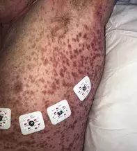
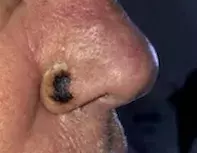
A 65-year-old man presents with a rapidly progressing diffuse petechial rash with associated malaise and fevers (Figure 1a). The rash was preceded by an insect bite on the right nasolabial fold that has evolved into an eschar (Figure 1b).
Differential diagnoses
Conditions to consider among the differential diagnoses include the following.
- Vasculitis. Vasculitides are a diverse group of diseases having a similar histopathological appearance with blood vessel wall inflammation. These conditions are classified by the predominant size of blood vessels affected (small, medium or large vessel).1 Cutaneous manifestations of vasculitis are common and usually indicative of small- and medium-sized vasculitides. The clinical hallmarks of small-vessel vasculitides include petechiae, slightly raised purpura (‘palpable purpura’), pustules, urticaria and splinter haemorrhages.1 There are many diverse causes of vasculitis, including infection (e.g. hepatitis B or C virus infection), connective tissue disease (e.g. systemic lupus erythematosus, rheumatoid arthritis), malignancy, inflammatory bowel disease and drugs (e.g. penicillins, retinoids); however, no precipitating factor is identified in 50% of cases.1 Vasculitides that commonly present with a petechial eruption include cutaneous small vessel vasculitis, Henoch-Schönlein purpura and secondary to connective tissue disease.1 Cutaneous small vessel vasculitis is a diagnosis of exclusion and is characterised by a vasculitic eruption consisting of purpura, petechiae and/or vesicles localised to the skin in areas susceptible to trauma, usually caused by drugs or infection.1 Blood tests and imaging are required to determine the pattern of organ involvement, while biopsy of an active lesion within 48 hours of onset is diagnostic to confirm the histopathological findings of fibrinoid necrosis and to ascertain any associated granulomatous inflammation or leukocytic infiltrate that may provide diagnostic clues to the size of the vessels involved.1
- Viral exanthem. Viral infections in adults can manifest with nonspecific bright macular morbilliform eruptions with associated petechiae.2 Involvement of the face, acral areas, buttocks and mucosa with associated enanthem, lymphadenopathy, fever and hepatomegaly usually differentiate a viral from a drug-induced eruption.2 Cytomegalovirus and human herpesvirus 6 or 7 can cause an infectious mononucleosis-like syndrome with an associated petechial eruption in immunocompetent individuals, the former often following a course of penicillin antibiotics in 30% of cases.2 Dengue fever, which is caused by the dengue virus, classically presents with fever, arthralgia, headache and retro-orbital pain with an associated rash. Erythema of the face develops within the first 48 hours of disease, followed by a morbilliform eruption with associated petechiae involving the trunk, face and limbs that spares palmar and plantar surfaces.2 Severe haemorrhagic dengue fever is often heralded by a morbilliform rash with extensive petechiae, purpura and ecchymosis, with subsequent risk of developing fatal disseminated intravascular coagulation.2
- Invasive meningococcal disease. Caused by the Gram-negative diplococcal bacteria Neisseria meningitidis, invasive meningococcal disease should always be considered in an unwell febrile patient with a rapidly progressing diffuse petechial eruption. Risk factors include age (infants and young adults), complement membrane attack complex deficiency, hyposplenia/asplenia and overcrowding.3 Early clinical signs of invasive meningococcal disease include nonspecific coryzal symptoms and fever, with associated signs of meningitis occurring between 12 and 15 hours of disease onset.3 The eruption can present initially as a blanching maculopapular eruption that may resemble a viral exanthem, later developing into the characteristic nonblanching, petechial and/or purpuric rash that coincides with meningococcal septicaemia.3 Prompt identification and initiation of intravenous antibiotics are required, as fulminant invasive meningococcal disease may cause long-term disability, such as amputation and neurological impairment, and has a mortality rate of between 5 to 20%.3
- Rickettsia. This is the correct diagnosis. Rickettsioses are caused by the transmission of Gram-negative obligate intracellular bacteria that are transmitted via infected arthropod vectors (e.g. Ixodid ticks) to cause disease in humans, with the spotted fever group infections caused by the genus Rickettsia.4 Inoculation occurs when there is inhalational or direct skin exposure to aerosolised faeces or transmission of saliva during a blood meal from an infected tick, with an incubation period of between three and 10 days, depending on the organism.4 Causative organisms vary geographically, thus a detailed travel history is warranted to determine potential exposures. There is an initial prodrome of malaise, followed by symptoms of meningism, arthralgia, myalgia, lymphadenopathy and persistent fevers.4 Patients may also recall a recent insect bite, which initially appears as an erythematous macule, and subsequently developing an eschar,4 as seen in Figure 1b. The cutaneous lesions initially appear within two days of exposure as erythematous macules that progressively become papular and then purpuric, with accompanying petechiae involving the distal extremities before centripedally involving the trunk and acral surfaces.4 Direct immunofluorescence of a skin lesion biopsy can demonstrate the rickettsial organism, as can serological testing, although these results usually are not expedient enough to influence the decision to commence antibiotic therapy.4
Management
A high index of clinical suspicion is required for rickettsial infections because delayed or untreated infections have mortality rates between 10 and 60%.4 It is relatively uncommon in Australia and mainly found on the east coast from Queensland to Victoria.5 Tetracyclines (e.g. doxycycline) are the first-line treatment for suspected rickettsial infections, and serological testing to confirm the diagnosis should not delay initiation of therapy.4 Failure to commence treatment within five days of onset is associated with a significant increase in mortality, with complications including disseminated intravascular coagulation and gastrointestinal haemorrhage.4
Outcome
For this patient, a provisional diagnosis of rickettsial infection was made and he was commenced on oral doxycycline 100mg twice daily. There was a rapid improvement in his clinical condition within 24 hours. Serological testing was performed prior to initiation of treatment and later returned a positive result.
References
1. Fiorentino DF. Cutaneous vasculitis. J Am Acad Dermatol 2003; 48: 311-340.
2. Korman AM, Alikhan A, Kaffenberger BH. Viral exanthems: an update on laboratory testing of the adult patient. J Am Acad Dermatol 2017; 76: 538-550.
3. Pace D, Pollard AJ. Meningococcal disease: clinical presentation and sequelae. Vaccine 2012; 30 Suppl 2: B3-B9.
4. Burnett JW. Rickettsioses: a review for the dermatologist. J Am Acad Dermatol 1980; 2: 359-373.
5. Sexton DJ, Dwyer B, Kemp R, Graves S. Spotted fever group rickettsial infections in Australia. Review Infect Dis 1991; 13: 876-886.
Parasitic diseases

