Peer Reviewed
Perspectives on dermoscopy
A dark mole with a woolly border
Abstract
The woolly border seen on dermoscopy of blue naevi is due to the presence of deeply pigmented spindle melanocytes blending with the surrounding skin within the dermis.
Key Points
Case presentation
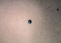
An 18-year-old man noticed a dark mole on his upper back (Figure 1), six months prior to consultation. He felt that the intensely pigmented mole (measuring 5 mm in diameter) had grown slowly in the interval. Dermoscopy revealed a symmetrical mole which had a homogeneous, structureless blue–black component in its centre, surrounded by a woolly border. The latter consisted of fine trails of blue–black pigment that were partially obscured by a light veil (Figure 2). Excision biopsy revealed a dermal infiltrate of deeply pigmented spindle-shaped melanocytes, which extended into the surrounding dermis but lacked nuclear atypia or mitoses (Figure 3).
Purchase the PDF version of this article
Already a subscriber? Login here.