Peer Reviewed
Perspectives on dermoscopy
A mole with a broken pigment network
Abstract
A broken and coarse pigment network may reflect the presence of a dermal component that has effaced the rete ridges.
Key Points
Case presentation
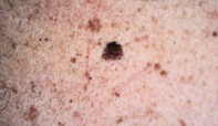
A 34-year-old man presented with a 1.2 x 0.9 cm, irregularly pigmented, flat mole of unknown duration on his upper back (Figure 1). Dermoscopy showed an asymmetrical lesion with a coarse pigment network which was darker at the superior edge. In many areas the pigment network was only partially represented, by linear and whorled remnants (Figure 2). The pale areas within the mole separating the network matched the colour of the surrounding skin. There was no evidence of scarring. Excision biopsy showed an epidermis with a prominent but irregular pigmented rete ridge system of differing lengths and a diffuse sheet of benign naevus cells in the underlying dermis (Figure 3).
Purchase the PDF version of this article
Already a subscriber? Login here.