Peer Reviewed
Perspectives on dermoscopy
A bleeding lesion with a villous surface
Abstract
Seborrhoeic keratoses often lose their characteristic dermatoscopic features, and pale villous projections may be the only clue to the correct diagnosis.
Key Points
Case presentation
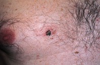
Over a six week period, a 64-year-old man noted an irritated lesion on his anterior chest (Figure 1). The lesion measured 8 mm in diameter and it bled. Dermoscopy revealed an elevated lesion with multiple pale villous projections that were partially obscured by a black eschar. The surrounding skin had a pink homogeneous hue, but there was no evident pigment network (Figure 2). Excision biopsy showed a markedly papillomatous epidermis covered by a loose stratum corneum containing locules of blood. The epidermis was hyperplastic but lacked evident atypia. There was a prominent lymphocytic infiltrate hugging the epidermal junction at the dermal interface (Figure 3).
Purchase the PDF version of this article
Already a subscriber? Login here.