A baby who drags one leg when crawling
Infant and newborn development
Should a late diagnosis of congenital hip dislocation be considered in the case of this 10-month-old baby?
Case presentation
History and examination
A 10-month-old baby girl presented with a history of dragging her left leg while crawling. She had been born at full term by cephalic presentation, and she had no family history of hip dysplasia.
Examination revealed an extra crease on the left leg with some shortening of the leg. Instability of the left hip was apparent, with a clunk on Ortolani’s manoeuvre.
Case presentation
History and examination
A 10-month-old baby girl presented with a history of dragging her left leg while crawling. She had been born at full term by cephalic presentation, and she had no family history of hip dysplasia.
Examination revealed an extra crease on the left leg with some shortening of the leg. Instability of the left hip was apparent, with a clunk on Ortolani's manoeuvre.
Diagnosis and treatment
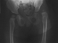
The patient was diagnosed with a dislocated hip, revealed at radiology (Figure 1). She underwent a closed reduction of the hip under general anaesthesia, and a percutaneous release of the abductor longus tendon was performed to facilitate hip abduction. She was fitted with a plaster hip spica for 10 weeks, followed by an abduction brace for three months. She walked at the age of 14 months.
X-rays later revealed some residual hip dysplasia with a steep acetabulum (Figure 2). When the child was 2.5 years old, an innominate osteotomy was performed to reduce the angle of the acetabulum and cover the femoral head (Figure 3). Internal fixation was performed using wires, and another plaster spica was applied. The latest x-rays, taken when she was 5 years old, reveal a normal hip joint (Figure 4).
Discussion
Neonatal examination and ultrasound have led to a significant decrease in late diagnoses of congenital hip dislocation, but cases do still occur. Risk factors include:
- female gender (the ratio of presentations in girls to boys is 10 to 1)
- breech presentation
- a family history of hip dysplasia
Clinical signs include a difference in the lengths of the left and right legs, as well as skinfold asymmetry with asymmetry of abduction. Barlow's and Ortolani's manoeuvres are provocative tests: Barlow's manoeuvre involves dislocationof the femoral head with flexion and abduction; Ortolani's manoeuvre is a reduction manoeuvre involving abduction and elevation of the greater trochanter.
Physiological laxity is common in the neonatal period, and ulltrasound is generally not recommended within the first two to three weeks. Any risk factors for hip dysplasia or clinical suspicion should lead to an ultrasound to allow this diagnosis to be made early. Treatment for cases that are diagnosed early generally involves a Pavlik harness, a dynamic brace that allows some movement but particularly restricts abduction. Cases that are diagnosed late can sometimes be treated with a closed reduction, as in the case described above, but are more likely to require an open reduction of the hip joint.
Key points
- Remember congenital hip dislocation in toddlers who have an abnormal gait or are slow to walk (that is, they delay walking until after 18 months of age).
- Have a high index of suspicion for hip dysplasia for a baby born by a breech presentation and any child with a family history of hip dysplasia. MT