Peer Reviewed
Perspectives on dermoscopy
Sessile pigmented lesions
Abstract
Dermoscopy is particularly useful in identifying small dark seborrhoeic keratoses.
Key Points
Case presentation
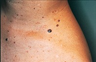
Over a 10-year period, a 52-year-old woman developed a series of pigmented papular lesions on her neck and upper trunk (Figure 1). Dermoscopic examination of the largest lesion showed a well demarcated symmetrical lesion with a smoky tan to light brown colour and numerous yellowish-white globules (Figure 2). The surrounding small papular lesions showed identical findings. A shave biopsy revealed a hypertrophic epidermis associated with small keratinocytes that enclosed multiple large keratin-filled pseudocysts (Figure 3).
Purchase the PDF version of this article
Already a subscriber? Login here.