A scar with striated pigmentation
Moles
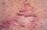
Case presentation
A 29-year-old man had had a benign mole removed from his upper back three years before the recent consultation. The mole had been removed by shave biopsy and cautery, but it had subsequently recurred. The site was then widely excised, producing a broad, wrinkled scar measuring 9 x 4 cm. Six months after surgery, streaks of pigment had appeared within the scar, producing a striated pattern (Figure 1). The pigmentary changes had remained stable and confined to the scar for at least two years.
Dermoscopy revealed an extensive, fine diameter pigment network producing a brush-like pattern and also scattered blue–black dots in the pale scar (Figure 2). Skin biopsy showed extensive dermal scarring with overlying epidermal hyperpigmentation and melanin pigment in the superficial dermis. No melanocytic proliferation or atypia were present (Figure 3).