Peer Reviewed
Perspectives on dermoscopy
A friable pigmented lesion
Abstract
Dermoscopy is useful in recognising regression in pigmented lesions, and close study of the clinical features as well as the histopathology may identify the primary lesion.
Key Points
Case presentation
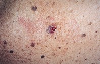
An 82-year-old man with multiple seborrhoeic keratoses developed an irritable pigmented lesion (measuring 1.3 cm x 1 cm) on his back (Figure 1). This had bled intermittently over a six-week period and had been itchy. Dermoscopy revealed an irregular pigmented lesion that had numerous blue–black dots giving a stippled pattern with pale scar-like areas adjacent to a raw bleeding surface (Figure 2). Excision biopsy showed an eroded epidermis with haemorrhage. The upper dermis contained abundant melanin pigment, lymphocytic inflammation and fibrosis. The adjacent epidermis was papillomatous and contained isolated keratin pseudocysts, but there was no melanocytic proliferation or atypia (Figure 3).
Purchase the PDF version of this article
Already a subscriber? Login here.