Peer Reviewed
Perspectives on dermoscopy
An irregular dark mole on the breast
Abstract
A slowly progressive, asymmetrical dark mole on a woman’s breast. Dermoscopy provides the clues to the diagnosis.
Key Points
Case presentation
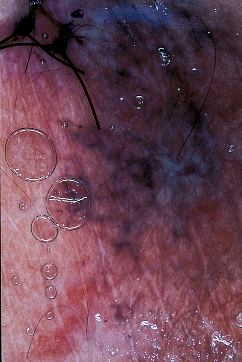
A 26-year-old woman had a longstanding, dark, flat, irregular pigmented lesion (1.3 x 0.9 cm) on her left breast. The pigmented lesion had been present since childhood and was associated with slow progressive growth. Dermatoscopy revealed mottled blue–black pigment in a retiform pattern covered by a patchy blue–grey veil. Skin biopsy showed an epidermis of normal thickness without junctional melanocytic nests or melanocytic proliferation. The underlying dermis had nests of uniform melanocytes (naevus cells) with sclerosis of collagen. There were pigmented nests of naevus cells in the superficial and mid dermis, but the deeper naevus cells were devoid of pigment.
Purchase the PDF version of this article
Already a subscriber? Login here.