Peer Reviewed
Perspectives on dermoscopy
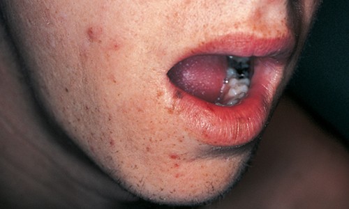
Pigmented lesion on lip
Abstract
The diagnosis of pigmented lesions is a daily challenge in general practice. Dermoscopy can provide extra clues, but requires significant expertise.
Key Points
Case presentation
Over a one-year period, a 27-year-old man noted a slowly enlarging pigmented lesion (0.4 cm diameter) on his right lower lip. His lips and face had numerous freckles. Dermoscopy showed a partially asymmetrical, light tan coloured patch with an irregular brown to grey pigmented network that was indistinct at its edges. Lip biopsy showed an epidermis that had a broad and blunt pigmented epidermal rete ridge system. Melanocytes were present as single cells and in normal numbers. The underlying dermis contained focal melanin pigment.
Purchase the PDF version of this article
Already a subscriber? Login here.