Peer Reviewed
Perspectives on dermoscopy
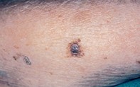
An asymmetrical mole with a blue–black centre
Abstract
Is it a naevus or a melanoma? Dermoscopy will help point towards the correct diagnosis.
Key Points
Case presentation
A 79-year-old woman presented with a 1.4 cm diameter lesion on her left forearm. The lesion was of unknown duration but had changed in colour and increased in size. Dermoscopic examination revealed an asymmetrical lesion with an irregular, variably pigmented, broken pigment network at the periphery. The central area had an irregular, blue–black, mottled colour partially covered by a blue–grey veil. The excision specimen showed an atrophic epidermis with focal asymmetrical junctional melanocytes and large nests of melanocytes that penetrated into the deep dermis and were associated with nuclear atypia.
Purchase the PDF version of this article
Already a subscriber? Login here.