Peer Reviewed
Feature Article Dermatology
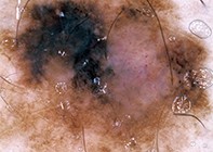
Melanoma: clinical features and early diagnostic techniques
Recent articles on:
Melanoma
Melanoma
Recent articles on:
Skin cancer
Skin cancer
Abstract
GPs are at the front line in the diagnosis of melanoma and are often required to assess pigmented lesions to determine if further investigation is warranted. Here is a review of the clinical features of melanoma and new techniques that have been developed to aid diagnosis.
Key Points
- Australia has the highest incidence of melanoma in the world, and GPs represent the front line of melanoma diagnosis.
- Assessment of melanoma risk factors enables GPs to identify high risk individuals and implement appropriate melanoma prevention and surveillance strategies.
- Dermoscopy and whole body photography add significant clinical information that may be used to identify new lesions or prevent unnecessary biopsy.
- Excisional biopsy with 2 mm margins is the preferred method for further investigation of suspicious pigmented lesions.
- Referral of patients to a multidisciplinary melanoma unit should be considered in complicated cases requiring specialist management.
- Computer assisted diagnosis is a new frontier in melanoma diagnosis. At present, this technology should be viewed with caution as it cannot reliably diagnose up to 20% of melanomas.
Purchase the PDF version of this article
Already a subscriber? Login here.