Peer Reviewed
Perspectives on dermoscopy
To BCC or not to BCC?
Abstract
The use of dermoscopy is described in two case presentations to differentiate melanocytic from other common skin lesions.
Key Points
Case presentations
Case 1
An elderly man with a history of solar keratoses and nonmelanoma skin cancer presented for a skin check. He had no specific skin concerns.
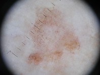
A dusky pink lesion was noted on the patient’s lateral right elbow (Figure 1a). On close inspection, the plaque was violaceous and ill defined, measuring approximately 9 x 7 mm. Although the patient and his wife were normally fairly observant of skin changes they were mostly unaware of the existence of the lesion, which suggested recent onset. It was asymptomatic.
Purchase the PDF version of this article
Already a subscriber? Login here.

