Peer Reviewed
Gastroenterology clinic
Noninvasive tests for assessing liver fibrosis
Abstract
Assessing the degree of liver fibrosis in patientswith chronic liver disease assists in management decisions and in determining prognosis. Given that liver biopsy is invasive and carries risks, there is much interest in less invasive technologies to stage liver disease.
Key Points
- Liver fibrosis develops via a common pathway as a result of liver injury and inflammation. In Australia, the most common causes of liver fibrosis are chronic hepatitis B, chronic hepatitis C, alcohol and nonalcoholic fatty liver disease. Interaction between risk factors for liver fibrosis is common, and the final stage of the process is the development of hepatic cirrhosis.
- The identification of patients with hepatic cirrhosis is crucial because all patients with cirrhosis should be checked for oesophageal varices and screened for the development of hepatocellular carcinoma (typically with a liver ultrasound examination every six months).
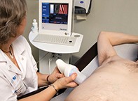
- Histological examination of a liver biopsy specimen remains the reference standard for the assessment of liver fibrosis. However, liver biopsy is invasive, potentially painful and associated with a small but definite mortality rate (0.01%). Also, the small sample obtained by biopsy increases the rate of sampling errors.
Remember
Purchase the PDF version of this article
Already a subscriber? Login here.