Thunderclap headache: when the risk of doing nothing is too high

A thunderclap headache is an uncommon presentation of headache but is important to recognise because it is often associated with serious vascular intracranial disorders, and should therefore be investigated urgently. Aneurysmal subarachnoid haemorrhage is the foremost consideration, and reversible cerebral vasoconstriction the next most common cause. The causes and diagnostic approach to patients with thunderclap headache are discussed.
- The cause of thunderclap headache should be considered to be aneurysmal subarachnoid haemorrhage until proven otherwise.
- The diagnostic work up involves urgent nonenhanced CT of the brain (with CT angiography if immediately available), then possibly a lumbar puncture, and then cerebral arterial and venous imaging with either MRI or CT if each preceding investigation has yielded a normal/negative result.
- Reversible cerebral vasoconstriction syndrome is usually characterised by recurrent thunderclap headaches and multifocal, multivessel segmental cerebral artery vasoconstriction that usually resolve within 12 weeks. It can be associated with neurological complications including intracerebral haemorrhage and cerebral ischaemic infarctions.
Picture credit: Juanmonino/iStockphoto.com
Model used for illustrative purposes only
A thunderclap headache is an uncommon presentation of headache but is important to recognise because it is often associated with serious vascular intracranial disorders, and should therefore be investigated urgently. Aneurysmal subarachnoid haemorrhage is the foremost consideration, and reversible cerebral vasoconstriction the next most common cause. The causes and diagnostic approach to patients with thunderclap headache are discussed.
Thunderclap headache is often associated with serious vascular intracranial disorders, particularly subarachnoid haemorrhage (SAH). The search for an underlying cause should be immediate and exhaustive. This article discusses the causes and diagnostic approach to patients with thunderclap headache.
Definitions and clinical presentation
Thunderclap headache is a high-intensity headache of abrupt onset, generally reaching maximum intensity in less than one minute, and lasting for five minutes or longer, according to The International Classification of Headache Disorders, 3rd edition beta.1 A practical diagnostic aid is to snap one’s fingers at the patient and ask: ‘Did it come on like this?’ The description ‘most severe ever headache’ does not, by itself, establish the diagnosis of thunderclap headache.
The history alone is often insufficient to determine the cause of the thunderclap headache with certainty. It is necessary to enquire about a prior history of headaches, recurrence of the thunderclap headache, the events around the onset of the headaches (sexual activity, physical activity, bath, cough, Valsalva manoeuvre), head and neck trauma, drugs used (serotonergic and sympathomimetic medications and illicit drugs) and pregnancy and postpartum status.
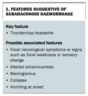
Certain symptoms and signs are more suggestive of certain underlying causes; for example, focal neurological symptoms or signs, altered consciousness, meningism, collapse or vomiting at onset would raise suspicion of a subarachnoid or even intracerebral haemorrhage (Box 1).
Causes
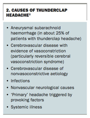
The list of causes of thunderclap headache is extensive, with the rare causes frequently reported in the medical literature as case reports only.2 The list can be divided into the following groups to assist with clinical reasoning (Box 2):
- aneurysmal SAH
- cerebrovascular disease with evidence of vasoconstriction – particularly reversible cerebral vasoconstriction syndrome
- cerebrovascular disease with no evidence of segmental arterial constriction, i.e. nonvasoconstrictive aetiology – includes intracranial haemorrhage (subdural, subarachnoid, intraparenchymal, epidural), ischaemic stroke, arterial dissection, cerebral venous sinus thrombosis, inflammatory arteriopathy and hypertensive encephalopathy
- infections – includes viral illness, rhinosinusitis, aseptic and bacterial meningitis
- nonvascular neurological causes – includes intracranial hypotension, pituitary apoplexy, cerebral neoplasms and colloid cyst of the third ventricle
- ‘primary’ headache triggered by provoking factors – primary cough headache, bath-related thunderclap headache, primary exercise headache, primary headache associated with sexual activity
- systemic illness – includes acute myocardial infarction, aortic dissection, phaeochromocytoma.
Diagnostic work up
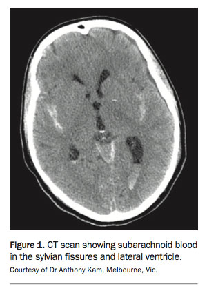
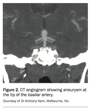
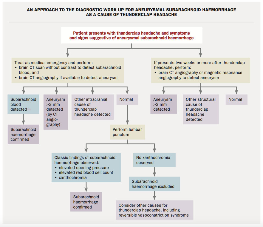
Following the history and physical examination, all patients with thunderclap headache should be managed as a medical emergency to avoid potentially catastrophic consequences from SAH and other intracranial causes of the headache. SAH is usually detected by brain CT imaging and the site of bleeding is often apparent on CT angiography (Figures 1 and 2).
If a patient presents two weeks or more after thunderclap headache, the top priority is to exclude an underlying aneurysm or other structural cause rather than to try to demonstrate subarachnoid blood. Proceeding directly to brain CT with CT angiography or MRI with magnetic resonance angiography would be appropriate.
The flowchart shows an approach to the diagnostic work up of a patient with thunderclap headache.
Brain CT scanning and angiography
The first investigation should be a brain CT scan to evaluate for SAH and other intracranial causes of thunderclap headache (Figure 1). If available immediately, CT angiography of the head should be performed at this time also (Figure 2).
The brain CT scan should be performed as soon as possible after the onset of the headache. CT brain imaging performed in the first six to 12 hours has a specificity of 98% and a sensitivity of close to 100% for detecting SAH. However, the sensitivity of CT to detect a SAH progressively declines as the interval between the headache and the CT imaging lengthens: it is about 85% to 95% on day 2, about 75% on day 3 and about 50% after day 5.3 The sensitivity is reduced in small volume bleeds, if the CT scan is not reviewed by an experienced reviewer or if the symptoms are atypical.
Lumbar puncture
A lumbar puncture is indicated if a brain CT scan or CT angiography does not reveal the aetiology of the thunderclap headache.
The classic findings of SAH on lumbar puncture are an elevated opening pressure and an elevated red blood cell count that does not diminish from cerebrospinal fluid tube one to tube four, and the presence of xanthochromia.
Xanthochromia represents haemoglobin degradation products. This indicates that blood has been in the cerebrospinal fluid for at least two hours. Xanthochromia is detected visually by comparing a vial of cerebrospinal fluid with a vial of plain water held side by side against a white background in bright light or by formal spectrophotometric analysis. Spectrophotometry detects bilirubin and is highly sensitive when the lumbar puncture is performed at least 12 hours after the SAH. Xanthochromia can last for two weeks or more.4
Further analysis of the cerebrospinal fluid includes cell counts and measurement of protein and glucose levels, and microscopy and culture to test for infections in the central nervous system.
MRI and angiography
Patients who have nondiagnostic brain CT scans and lumbar puncture results should be further evaluated with contrast-enhanced brain MRI and noninvasive vascular imaging of the head and neck (magnetic resonance angiography or CT angiography). This imaging can detect arterial dissections and vasoconstriction syndromes.
CT venography and magnetic resonance venography
Venous sinus imaging via CT or magnetic resonance venography is indicated if clinical suspicion for a cerebral venous thrombosis is high.
Subarachnoid haemorrhage
About 70% of patients with SAH present with headaches as the main symptom, and about 50% of these present with thunderclap headache. SAH is found in up to 25% of patients with thunderclap headache. Aneursymal SAH is therefore the diagnosis of foremost consideration in this scenario.
The brain CT Scan and/or the lumbar puncture would be expected to yield a positive result in patients with SAH. Magnetic resonance angiography or CT angiography will often detect aneurysms, but only those larger than 3 mm. Formal four-vessel catheter angiography (cerebral angiography) is the gold standard to determine the site and morphology of the aneurysm and to guide treatment (angiography with a catheter also offers the possibility of coiling the aneurysm).
Treatment of SAH is highly specialised and patients require management in a well-equipped expert neurosurgical unit. Management includes supportive care and avoidance of vasoconstriction, which may produce cerebral ischaemia. The cornerstone of treatment is to prevent further bleeding from the cause (usually an aneurysm). This requires surgical securing of the aneurysm (by clipping) or endovascular treatment (with coils and similar devices).
Reversible cerebral vasoconstriction syndrome
A greater understanding of reversible cerebral vasoconstriction syndrome (RCVS) has emerged in the past few years because of the wider availability of relatively noninvasive technologies to assess the cerebral vasculature. This syndrome has been given various labels (e.g. Call–Fleming syndrome, benign angiopathy of the central nervous system, postpartum angiopathy and migrainous vasospasm), and can occasionally be confused with cerebral vasculitis because of overlapping angiographic and clinical features. The term ‘reversible’ refers to the intracranial artery vasoconstriction and not the possible permanent neurological deficits secondary to complications including intracerebral haemorrhage and cerebral ischaemic infarctions. RCVS may occur at any age, but many patients are young. Although most patients recover fully, a minority suffer permanent cerebral ischaemic damage.
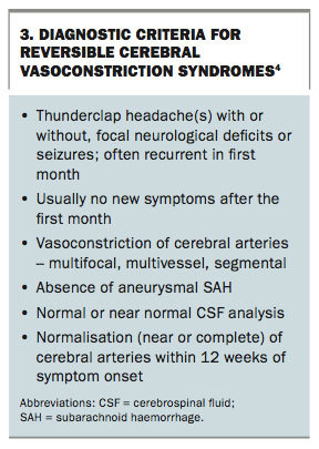
The diagnostic criteria for RCVS include (Box 3):5
- thunderclap headache(s) with or without focal neurological deficits or seizures
- often recurrent episodes of thunderclap headache during the first month but usually no new symptoms appearing after this time
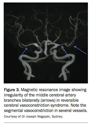
- multifocal, multivessel, segmental vasoconstriction of cerebral arteries. The vessels involved may be main trunks of vessels such as middle or posterior cerebral arteries but are often more peripheral branches. The changes may be obvious but are sometimes subtle and only detected when the radiologist is alerted to this as a possible diagnosis (Figure 3)
- absence of aneurysmal SAH
- normal or near normal cerebrospinal fluid analysis
- complete or substantial normalisation of cerebral arteries within 12 weeks of symptom onset.
RCVS can occur without an identifiable precipitating factor, during pregnancy or the puerperium period and as an idiosyncratic response to certain medications (selective serotonin reuptake inhibitors and nasal decongestants have been implicated) or illicit drugs (either acute or chronic use) and in the setting of catecholamine-secreting tumours. It can be provoked by sexual activity, exertion or Valsalva manoeuvres, emotions and bathing/showering.
It is usual to have multiple thunderclap headaches over one to four weeks, sometimes with a baseline lingering headache in between. A small percentage of patients (5%) have recurrences after this period.
Lumbar puncture analysis is typically normal in patients with RCVS. A significantly abnormal cerebrospinal fluid analysis (with pleocytosis) should prompt consideration of the differential diagnosis of primary angiitis of the central nervous system, a rare condition, or other forms of cerebral vasculitis. (Radiologists often raise the question of cerebral vasculitis in RCVS cases but RCVS is much more common than cerebral vasculitis, especially when the presentation is thunderclap headache).
Brain CT and MRI scans are normal in uncomplicated RCVS but can reveal abnormalities otherwise. Early complications, mainly occurring in the first week of symptoms, include cortical SAH (usually very localised within a sulcus as opposed to the widespread SAH typically seen after ruptured aneurysm), intracerebral haemorrhage and cerebral vasogenic oedema, as seen in the posterior reversible encephalopathy syndrome. Ischaemic events, including transient ischaemic attacks and cerebral infarction, occur mainly in the second week. In one study, these complications occurred at rates of cortical SAH, 22%; intracerebral haemorrhage, 6%; cerebral vasogenic oedema, 9%; transient ischaemic attacks, 16%; and cerebral infarction, 4%.6
Angiography (magnetic resonance or CT) in RCVS will reveal multifocal vasoconstriction of multiple intracranial arteries that is maximal at about two to three weeks after symptom onset. The pattern is one of distal vasoconstriction initially, which may then move more proximally in the first several weeks after symptom onset, and then resolves (is ‘reversible’). Normal vasculature on initial angiography should prompt repeat vascular imaging in a few weeks to search for vasoconstriction.
Treatment for RCVS is still guided by observational and anecdotal experience. Identifying the precipitating factors or disease guides treatment (e.g. discontinuation of vasoactive drugs, resection of catecholamine-secreting tumours). There is probably a role for the off-label use of calcium channel blockers (nimodipine or verapamil) as first-line treatment. Short-term use of high-dose glucocorticoids is controversial: it had been reported as effective based on reversal of vasoconstriction in experiments but has been accused of worsening clinical outcomes.7 Some argue that simple observation with follow-up imaging may be reasonable as the disease course is self-limiting.5 Avoiding further vasoconstrictors, such as ergots and triptans, would seem prudent.
Primary headaches
Although there are diagnostic criteria in The International Classification of Headache Disorders, 3rd edition beta, for primary thunderclap headache, the evidence that this exists as a primary disorder is poor and this diagnosis should only be made with caution and as a last resort.1
Other primary headaches that are provoked (i.e. primary cough headache, primary exertional headache and primary headache associated with sexual activity) have a number of possible secondary causes as well, which must first be ruled out.
There is now increasing evidence that ‘primary thunderclap headache’, some of the primary headaches that are provoked and RCVS are part of the same spectrum. For example, many cases of headache associated with sexual activity (so-called ‘benign sex headache’) were shown on careful angiography (CT or magnetic resonance) to have cerebral vasoconstriction as expected in RCVS; these cases were clinically indistinguishable from those in whom cerebral vasoconstriction was not found.8 Furthermore, these conditions often follow a similar time course, and patients with headache associated with sexual activity will be at risk of recurrent episodes for four to six weeks but may then never experience them again, just like those with proven RCVS.
Conclusion
A thunderclap headache is an uncommon presentation of headache but is important to recognise because it should prompt urgent medical review and investigation as it is often associated with serious vascular intracranial disorders. Aneurysmal SAH is the foremost consideration. Understanding of RCVS is expanding and this is probably the next most common cause for thunderclap headache. The clinical importance of these disorders highlights the need for a precise history of how a headache develops and evolves. Any instantaneous onset headache requires urgent and careful evaluation. MT
COMPETING INTERESTS: Dr Cheng: None. Associate Professor Stark has received lecture fees and acted on advisory boards for Allergan, Novartis and SciGen.
References
- Headache Classification Committee of the International Headache Society. The International Classification of Headache Disorders, 3rd edition (beta version). Cephalagia 2013; 33: 629-808.
- Devenney E, Neale H, Forbes RB. A systematic review of causes of sudden and severe headache (thunderclap headache): should lists be evidence based? J Headache Pain 2014; 15: 49.
- Beckes D, Rinkel G, Kemperman H, Linn FH, Vergouwen MD. Time-dependent test characteristics of head computed tomography in patients suspected of nontraumatic subarachnoid haemorrhage. Stroke 2012; 43: 2115-2119.
- Vermeulen M, Hasan D, Blijenberg BG, Hijdra A, Van Gijn J. Xanthochromia after subarachnoid haemorrhage needs no revisitation. J Neurol Neurosurg Psychiatry 1989; 52: 826-828.
- Calabrese L, Dodick D, Schwedt T, Singhal AB. Narrative review: reversible cerebral vasoconstriction syndromes. Ann Intern Med 2007; 146: 34-44.
- Ducros A, Boulkobza M, Porcher R, Sarov M, Valade D, Bousser MG. The clinical and radiological spectrum of reversible cerebral vasoconstriction syndrome. A prospective series of 67 patients. Brain 2007; 130: 3091-3101.
- Singhal AB, Hajj-Ali RA, Topcuoglu MA, et al. Reversible cerebral vasoconstriction syndromes: analysis of 139 cases. Arch Neurol 2011; 68: 1005-1012.
- Yeh Y, Fuh JSC, Wang SJ. Clinical features, imaging findings and outcomes of headache associated with sexual activity. Cephalalgia 2010; 30: 1329-1335.