Peer Reviewed
Perspectives on dermoscopy
A pigmented lesion with a white patch
Recent articles on:
Melanoma
Melanoma
Abstract
Pale structureless areas and pale areas containing multiple blue–black dots correspond to areas of tumour regression. Asymmetrical and patchy tumour regression is seen particularly in melanomas.
Key Points
Case presentation
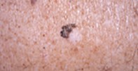
A 75-year-old man with extensive sun-damaged skin presented with an irregularly pigmented lesion of unknown duration, measuring 0.8 cm x 1.0 cm, on his back. The pigmented portion formed a crescent around a white patch (Figure 1). Dermoscopy revealed an irregularly pigmented lesion with a coarse and broken pigment network and streams of pigment at the edge. There were multiple blue–black dots and scattered pale areas within the pigmented segment. The large white patch appeared structureless (Figure 2). The excision specimen showed an atrophic epidermis with confluent atypical melanocytes present along the junctional zone together with extensive dermal fibrosis containing clumps of melanin pigment and lymphocytes (Figure 3).
Purchase the PDF version of this article
Already a subscriber? Login here.