Peer Reviewed
Perspectives on dermoscopy
A recurrent black lesion
Recent articles on:
Skin cancer
Skin cancer
Abstract
Some pigmented basal cell carcinomas may be difficult to distinguish from melanomas on dermoscopy.
Key Points
Case presentation
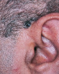
A 52-year-old man developed a progressively enlarging pigmented lesion, measuring 3.0 cm x 1.0 cm, above his left ear (Figure 1). Six years previously, a dark pigmented nodule had been removed adjacent to this site. Dermoscopy revealed an asymmetrical black lesion with an irregular border shaped vaguely like maple leaves. There were streams of pigment at the edges and focal blue–black dots. The bulk of the lesion showed structureless black pigmentation with a hazy blue–white veil (Figure 2). Excision biopsy of the lesion showed large basaloid lobules of tumour, which penetrated into the deep dermis and were associated with patchy melanin pigment (Figure 3).
Purchase the PDF version of this article
Already a subscriber? Login here.