Fitting the fits. Common paediatric epilepsy syndromes
Epilepsy affects 1 to 2% of the population, including children. Revised epilepsy classifications have been published recently, aiming to clarify the diagnosis of epilepsy syndromes. GPs are encouraged to use the new classifications to better diagnose and manage paediatric patients with epilepsy and help inform their anxious families.
Epilepsy affects 1 to 2% of the population, including children. Paediatric epilepsy is anxiety-provoking for families and medical practitioners. Although seizures are always a cause for concern, there are a number of common epilepsy conditions that are self-limiting and often well-controlled.
Revised classifications for paediatric epilepsy, published by the International League Against Epilepsy (ILAE) in 2017, are designed to improve the diagnosis of epileptic syndromes, with new terminologies for seizure classifications.1 The ILAE Seizure Classification presents three levels of diagnosis: seizure type, epilepsy type and epilepsy syndrome. The aetiology of a patient’s seizures is considered at each level of classification.1
Understanding seizure classifications, common paediatric epilepsy syndromes, prognosis and investigative approaches in the context of the updated ILAE classifications can aid in the understanding of likely outcomes and assist in the management of paediatric patients with epilepsy and their families.
Seizure classification
To better understand what is occurring in infants and children with epilepsy, it is important to first understand the type of seizure occurring. GPs are encouraged to use the new ILAE terminology as it is designed to better distinguish between seizure types and allow better recognition of epilepsy syndromes.
The first thing to determine in a paediatric patient presenting with seizure-like symptoms is whether an episode or repeated episodes are consistent with seizure, as many nonepileptic events (e.g. vasovagal episodes) masquerade as seizures. According to the new classification system, the first step of diagnosis is confirmation of a seizure type. Following this, the epilepsy type is further classified into generalised seizures and focal seizures (although in some patients the seizure type remains unknown). Generalised seizures include generalised tonic-clonic seizures, absences, myoclonus, atonic, tonic and clonic seizures. In older terminology, absence seizures were described as petit mal seizures, whereas generalised tonic-clonic seizures were known as grand mal seizures (‘little bad’ and ‘big bad’ seizures, respectively).
Focal seizures were previously known as partial seizures. Focal seizures begin in one region of the brain, with stereotyped manifestations resulting from the location of the seizure onset and spread. Previously, depending on whether consciousness was affected, focal seizures were further divided into simple partial (no impairment of consciousness) or complex (dyscognitive or impaired cognition/consciousness). Focal seizures are now described as being with or without impairment of awareness/consciousness.
A common error in diagnosing seizures is to categorise all episodes with staring and unresponsiveness as petit mal seizures, without recognising the distinction between focal seizures and absences. This distinction is important in determining seizure aetiology, treatment and prognosis. Features to consider when assessing patients with seizures are shown in Box 1. The differential diagnosis of seizures in childhood is shown in Box 2. Common imitators of seizures vary by age group.
Common epilepsy syndromes
Childhood absence epilepsy
Childhood absence epilepsy (CAE) is characterised by the occurrence of numerous seizures a day. These seizures manifest as interruption of concentration, speech and activity, resulting from a brief loss of awareness. Automatisms (e.g. eye blinking and lip smacking) can be present. These seizures, previously classified as petit mal, account for around 10% of childhood epilepsies. Onset is typically around the age of 5 to 6 years but ranges from 2 to 10 years. Children with CAE will usually only have absence seizures, and the seizures usually remit by adolescence (about age 12 years). Diagnosis is based on clinical description and an EEG, which typically shows a 3 Hz spike and wave pattern at the time of an event, often brought on by hyperventilation. Neuroimaging is not usually required.
Medication should be considered for children with CAE because ongoing events can lead to problems attending to classwork or to safety issues (e.g. crossing the road). Typical medications used in the management of CAE include ethosuximide, sodium valproate and lamotrigine, with a randomised controlled trial recommending ethosuximide as first-line treatment, balancing side effects and efficacy.2
Most children with absence seizures will become seizure-free with time, but some patients will go on to develop other epilepsy syndromes, usually other generalised epilepsies. Ethosuximide only treats absence seizures; it does not protect against other seizure types, so if these occur a change in medication may be needed, with valproate or lamotrigine often considered. Possible predictors of evolution to other epilepsy syndromes include later age of onset or atypical EEG features.
Absence seizures can be confused with focal seizures (previously called complex partial seizures). Absences are often brief (5 to 10 seconds) with an immediate return to the usual levels of responsiveness, whereas focal seizures are often longer and are followed by behavioural change and sleepiness. An EEG may show focal (i.e. localised) activity rather than generalised activity, and an MRI is warranted to look for any underlying structural abnormality (Figure 1 and Figure 2). Some focal seizures can evolve into generalised tonic-clonic seizures or can have ictal posturing or postictal hemiparesis.
Childhood epilepsy with centrotemporal spikes
Common focal epilepsies in childhood include childhood epilepsy with centrotemporal spikes (previously known as benign rolandic epilepsy or benign epilepsy of childhood with centrotemporal spikes). This is the most common school-age epilepsy (15 to 25% of patients) and is said to peak at 8 to 9 years of age. Seizures are infrequent, and 10% of children diagnosed with childhood epilepsy and exhibiting centrotemporal spikes have only one seizure. Seizures often occur while the child is asleep (75%), although they can occur during the day. The child typically makes a groaning or gagging sound at the start of a seizure because of the involvement of the mouth and face, and may be aware but unable to speak. These seizures can remain focal or can evolve into clonic movements on one side of the body that then spread bilaterally.
An EEG typically shows focal activity over bilateral centrotemporal regions, which occurs independently and becomes more frequent and obvious during sleep. If an initial EEG is normal but an epilepsy syndrome is suspected, an EEG after sleep deprivation may be useful to bring out the activity during sleep. Clinical correlation is important to ensure a seizure is consistent with the ictal description because the same EEG pattern can occur in about 2 to 3% of children without the occurrence of seizures. Neuroimaging is not required unless there are atypical features on the EEG or clinically. Medication should be considered if seizures are multiple and particularly if they are frequent. Carbamazepine, sodium valproate or levetiracetam are common choices. Seizures should remit in adolescence. If seizures reoccur after this, an alternative diagnosis should be considered and the patient re-evaluated.
Panayiotopoulos syndrome
Panayiotopoulos syndrome is characterised by autonomic seizures that occur in early childhood (starting from 3 to 6 years of age) and accounts for 2 to 5% of childhood epilepsy. Events are often prolonged and last for 20 or 30 minutes. Autonomic features in the seizure can include profound colour change (pallor or cyanosis), vomiting, hypersalivation and incontinence. In some patients, there is eye deviation or unilateral clonus, and in 50% there is progression to convulsive seizures. An EEG can show occipital or multifocal activity, often with a posterior emphasis. Events are usually few and mostly occur in sleep. MRI should be considered if focal epilepsy remains a differential diagnosis, as focal pathology can cause similar seizures. When medications are used, carbamazepine and sodium valproate are usually considered initially.
Febrile convulsions
Febrile seizures occur in around 2 to 4% of children in the general population. Children are susceptible to febrile seizures from 6 months of age and can experience multiple seizure events up to the age of 6 years; however, many will experience only a single event. Most children with febrile convulsions do not go on to develop epilepsy, and are at no greater risk of developing epilepsy than the general population. A family history of febrile convulsions is reported in around 20% of cases of children with febrile convulsions.
Febrile convulsions can be classified as simple or complex; simple febrile convulsions are shorter (less than 15 minutes), with tonic-clonic movements and occur as a single event in the illness. Children with complex febrile convulsions are more likely to go on to develop afebrile seizures and epilepsy, although most are seizure- free in the long term. In diagnosing febrile convulsions, seizures must be accompanied by a fever – if no fever is present then it is not a febrile convulsion. Temperatures are usually high (e.g. 39oC) rather than being close to normal. Clinicians must be careful not to overlook a serious infection (e.g. meningitis), as this can also cause seizures with fever.
Prognosis for children with epilepsy syndromes
Some epilepsy syndromes occurring in childhood are more likely to continue into adulthood. This prognostication is an important reason for accurate diagnosis of epilepsy syndromes. Focal seizures occurring outside of the benign childhood syndromes can continue, particularly if there is an underlying cause (e.g. cerebral palsy or scarring from trauma) that may be found on MRI. Some common generalised seizure syndromes are likely to continue into young adulthood, including juvenile absence epilepsy and juvenile myoclonic epilepsy (JME). For 75% of patients with JME, the epilepsy begins in adolescence (age 12 to 18 years) and requires ongoing treatment into young adulthood. Myoclonus soon after waking, combined with generalised tonic-clonic seizures and, occasionally, absences, are the common seizure types in JME.
Investigations for paediatric epilepsy
Both EEG and MRI are common investigations for epilepsy. There is consensus among specialists that EEG is not required if the patient’s history is classical for an alternative diagnosis, such as breath holding, and many specialist centres do not encourage EEG for simple febrile convulsions. Warning signs for more urgent referral or neuroimaging are outlined in Box 3.
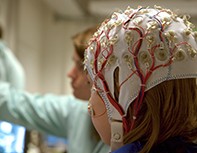
A normal EEG usually takes around 20 minutes to record and is a helpful start in investigating possible seizures, but does not have absolute sensitivity. The pickup rate of epileptiform activity can be increased by performing an EEG close to the time of a seizure (within 24 hours) or after sleep deprivation. The amount of sleep deprivation depends on the age of the child, with older children requiring longer sleep deprivation. It is important that the child stay awake while travelling for the EEG on the day. The electrographic signature on an EEG can help determine the epilepsy syndrome, in combination with the clinical features. Provoking manoeuvres such as hyperventilation and photic stimulation should be used in every EEG where feasible to bring out epileptiform activity.
An MRI scan can be helpful in looking for structural abnormalities and is particularly important in assessing focal epilepsies, which can be caused by underlying pathology. New onset seizures can occur in children with longstanding pathologies including underlying scarring from trauma or cerebral palsy, underlying areas of focal cortical dysplasia or low-grade tumours. However, most children with new onset focal seizures have no abnormalities on MRI. Children aged 5 years and older can usually tolerate an MRI with sufficient preparation. It may help to warn them of the noise in the MRI machine, and reassure them that it is not going to hurt them. Younger children usually need an anaesthetic to tolerate an MRI scan.
Paediatric epilepsies are usually well-controlled by medication and do not cause other problems. Points to consider when medication is not effective are shown in Box 4. Practice points on managing epilepsy in children are outlined in Box 5.
Conclusion
Improved knowledge of common paediatric epilepsy syndromes is helpful in managing patients and in explaining what is occurring to their anxious families. Many children with epilepsy will settle with time, and will not go on to have further seizures in adulthood. MT