Peer Reviewed
Perspectives on dermoscopy
When the hue is blue
Recent articles on:
Melanoma
Melanoma
Recent articles on:
Skin cancer
Skin cancer
Abstract
Clinicians need to be mindful of certain limitations of dermoscopy. These include a predominantly blue colour in lesions, which is relatively nonspecific when observed dermoscopically.
Key Points
Case presentations
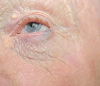
Case 1
A 75-year-old man presented for assessment of a solitary blue nodule arising from the margin of the left lateral lower eyelid. He reported that the lesion had been present and stable for ‘months’. The nodule was fixed, rather than fluctuating, and it was asymptomatic. The patient had a history of significant recreational sun exposure and multiple nonmelanoma skin cancers.
On examination, the nodule was firm and blue and measured 5 x 3 mm. The overlying eyelashes were intact. There were background changes of dermatoheliosis (photoageing) and a number of yellowish periocular milial cysts.
Purchase the PDF version of this article
Already a subscriber? Login here.