What’s the diagnosis?
A photodistributed rash in a malnourished patient


Case presentation
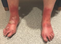
A 48-year-old woman presents with a three-week history of pruritic erythema on the dorsal aspect of her forearms and hands (Figures 1a and b) and feet (Figure 2). The eruption is sharply marginated, hyperpigmented and hyperkeratotic. Over the past 24 hours, she has developed bilateral ankle and foot oedema surmounted by tense bullae (Figure 2). The eruption is photodistributed; there is no rash on any other area of her skin.
The patient is otherwise well. She explains that she underwent gastric sleeve surgery three months previously. She became homeless recently and is living in a refuge.
Differential diagnoses
Conditions to consider among the differential diagnoses include the following.
- Porphyria. Porphyrias are a group of rare inherited metabolic disorders caused by altered activities of enzymes within the haem biosynthetic pathway. A common feature of all porphyrias is the accumulation of porphyrins or porphyrin precursors in the body, which results in photosensitivity. The cutaneous porphyrias can be divided into those causing chronic, blistering photosensitivity and those causing acute, nonblistering photosensitivity. The most common porphyria with an onset in patients of middle age is porphyria cutanea tarda. Patients are usually alcohol dependent and present with photosensitivity of the face, hyperpigmentation and scarring blisters on the dorsa of the hands.
- Polymorphic light eruption. This eruption usually presents as a pruritic rash in sun-exposed areas hours to days after sun exposure and persists for several days before subsiding. It is common and has a wide range of severity. A polymorphic light eruption is a recurrent, delayed-onset abnormal reaction to sunlight that resolves without scarring. It is not usually bullous.
- Chronic actinic dermatitis. This chronic dermatitis mainly affects men over the age of 50 years. It is characterised by the insidious onset of severely itchy, erythematous, inflamed and thickened dry skin, predominantly affecting photo-exposed sites, in association with objective evidence of photosensitivity. Bullae are not usually seen in this condition.
- Photosensitive drug eruption. Drug-induced photosensitivity occurs when certain photosensitising medications cause an unexpected sunburn in response to minimal light exposure or a dermatitis. Most drug-induced photosensitivity reactions are phototoxic rather than photoallergic. Phototoxic reactions appear as exaggerated sunburn, whereas photoallergy is more likely to be dermatitic. The phototoxic reaction usually evolves within minutes to hours of sun exposure and is restricted to exposed skin (face, neck, arms, backs of hands and often lower legs and feet). Common medications that cause photosensitive reactions include doxycycline, isotretinoin and thiazide diuretics. The patient described above was not taking any photosensitising medications.
- Acute cutaneous lupus. Cutaneous lupus is an inflammatory disorder that can be subdivided into a localised form in which lesions are confined to the face above the chin, the scalp and the ears and a disseminated form in which lesions also occur elsewhere on the body. It is characterised by well defined, red, scaly patches of variable size, which heal with atrophy, scarring and pigmentary changes. It can be bullous, but this is very rare. Patients are usually unwell with malaise and fever.
- Pellagra. This is the correct diagnosis. Niacin (nicotinic acid and nicotinamide) is an essential nutrient involved in the synthesis and metabolism of carbohydrates, fatty acids and proteins. Niacin deficiency causes pellagra, which is characterised by a pigmented dermatitis, diarrhoea and, when advanced, dementia. The dermatitis is photosensitive and typically located in sun-exposed areas. Other clinical findings can include angular stomatitis, cheilitis, glossitis and oral ulcers. In the Western world, pellagra is very rare and usually found in patients who have a very poor diet or are alcohol dependent. It has been associated with carcinoid syndrome.
Management
Pellagra is progressive and can be fatal if not treated. Treatment involves nicotinamide 500 mg/day, to which there is a dramatic response. When present, neuropsychiatric symptoms improve within the first 24 to 48 hours of treatment whereas cutaneous disease may take a few weeks to subside. Nicotinic acid is often avoided because it can cause headaches and flushing.
In this patient’s case, pellagra appeared to be a consequence of prior gastric surgery in combination with a poor diet. As the niacin deficiency was a result of decreased dietary intake, a multidisciplinary approach was taken to help resolve the underlying social issues. Her rash resolved rapidly, within a few days of commencing a normal diet.

