What’s the diagnosis?
A man with erythematous and swollen hands


Case presentation
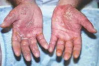
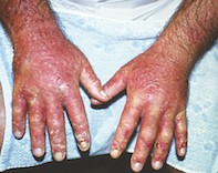
A 45-year-old man presents to the emergency department with a two-day history of progressive erythema and swelling affecting both hands (Figures 1a and b). He complains that they are painful and itchy.
The patient has a long history of a chronic scaly rash affecting the palms of both hands but has not had a similar acute rash before. He has been using an antiseptic hand wash for the past seven days because of increasing scale on his palms.
On examination, an erythematous vesiculobullous eruption is observed over the palmar and dorsal surfaces of both the patient’s hands. The rash extends to the dorsal forearms. Apart from lichenified, scaly skin over the extensor surfaces of his elbows, his skin is normal.
Differential diagnoses
Conditions to consider among the differential diagnoses include the following.
- Atopic dermatitis. This is a very common skin condition that manifests with intense itch. Atopic dermatitis is typically associated with a history of atopy such as hay fever or asthma. Acute lesions are characterised by erythematous papules that can weep and become vesicular when infected or very acute, which is similar to the morphology of this patient’s rash. However, acute atopic dermatitis does not involve the palms.
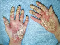
- Psoriasis. There is a wide spectrum of disease in psoriasis. Most commonly, psoriasis affects the extensor surfaces of the arms and legs. Scalp, palmoplantar and nail psoriasis are also common. In mild cases, these can be the only sites affected. The morphology of the lesions is typically well defined erythematous plaques covered in overlying white scale; however, sterile pustules, rather than vesicles, can occur (Figure 2). For this patient, the initial scaly rash on his palms may have been psoriasis but this is not the cause of the presenting eruption, which is characterised by acute blistering that is not consistent with psoriasis.
- Porphyria. The porphyrias are a group of acquired or congenital metabolic diseases that result in cutaneous manifestations. They are caused by dysfunction of the enzymes associated with haem biosynthesis – the resulting porphyrins accumulate in the skin and cause photosensitivity. Porphyria cutanea tarda is the most common porphyria and can present with blistering on the dorsal, but not palmar, surfaces of the hands. However, this is not an acute event, the blisters are not confluent and symptoms are minimal.
- Irritant contact dermatitis. Irritant contact dermatitis accounts for 80% of all contact dermatitis. It is particularly common among people with occupations that require frequent handwashing or repetitive handling of irritating substances. The resulting rash is a poorly demarcated erythematous eruption that can be both itchy and painful due to fissuring. In this patient’s case, the antiseptic wash he has been using could be implicated in irritant contact dermatitis, but such an intense reaction would not be expected over a short period of time. Additionally, irritant contact dermatitis does not extend beyond the area of skin that has been in contact with the irritant and would be unlikely to spread to the forearms if he used the product for only his hands.
- Infection. Staphylococcal and streptococcal infections can occur anywhere on the skin. The rash of cellulitis, which is usually due to group A Streptococcus pyogenes, manifests as a well-demarcated spreading erythema associated with oedema. Vesicles and bullae may be present. The rash tends to be painful rather than itchy and the patient is usually febrile and unwell. In this case, an alternative diagnosis is more likely, in light of the symmetry of the eruption (like ‘bilateral cellulitis’ of the lower limbs, which is almost always dermatitis with or without superinfection). As in all rashes with blistering and fissuring, there is a significant chance of secondary infection with Staphylococcus aureus.
- Allergic contact dermatitis. This is the correct diagnosis. Allergic contact dermatitis is a type IV cell-mediated immune response that presents after re-exposure to an allergen in an individual who has been previously sensitised. Acute episodes result in an erythematous, vesiculobullous eruption and extend beyond the area in contact with the allergen. Chronic exposure to an allergen can eventually be similar to chronic irritant contact dermatitis with eventual lichenification and scale. Common allergens include fragrances, metals (especially nickel) and plants (e.g. Robyn Gordon grevillea, poison oak). Antiseptics are also common allergens. Allergic contact dermatitis is common among nurses, dentists, hairdressers and workers in other occupations who handle rubber, nickel or epoxy resin.
Investigations
Identification of allergic contact dermatitis is mainly based on clinical suspicion, supported by resolution on withdrawal of the suspected allergen. Swabs of the rash should be performed to identify any secondary bacterial infection. Patch testing with the suspected allergen and the Australian Baseline Series is required to confirm the diagnosis.1
Management
Management of allergic contact dermatitis is twofold. Firstly, the acute rash is managed with withdrawal of the likely allergen and treatment of symptoms. Potent topical corticosteroids, such as betamethasone dipropionate ointment, are used with wet dressings to reduce itch and inflammation. A short course of oral prednisone 25 mg daily is also effective. Oral antibiotics may be required to treat any secondary infection. After the acute episode, avoidance of irritants and use of emollients is required for up to three months because the injured skin can remain vulnerable.
Outcome
For this patient, the eruption was treated with a two-week course of oral prednisone, topical betamethasone dipropionate 0.05% twice daily under wet dressings and cephalexin 500 mg four times daily for five days. After two weeks, he had recovered with desquamation and subsequent dryness and sensitivity which was managed with emollients and soap avoidance. He was advised to continue this for three months.
Patch testing was performed and the antiseptic chloroxylenol identified as the culprit. Allergen avoidance was advised to prevent further episodes. In some patients, especially those with occupational exposure to the allergen, this may prove very difficult, but fortunately this patient was easily able to avoid the product.
Reference
1. Toholka R, Wang YS, Tate B, et al. The first Australian Baseline Series: recommendations for patch testing in suspected contact dermatitis. Australas J Dermatol 2015; 56: 107-115.
Dermatitis

