What’s the diagnosis?
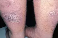
Cobblestone plaques on the legs


Kaposi’s sarcoma usually presents as multicentric violaceous plaques and nodules which may be concentrated over the lower limbs. Kaposi’s sarcoma may also be preceded by lower limb oedema, but is more rapidly progressive than the indolent lesions in this case. Skin biopsy shows ill-formed vessels dissecting the collagen bundles. The vessels are lined by atypical endothelial cells which may be associated with spindle cell differentiation. Viral stain for HHV8 can be demonstrated in the atypical endothelial cells in skin sections.
Cutaneous granulomas, particularly sarcoidosis, may also present as violaceous papules and nodules. Skin biopsy shows multiple granulomas consisting of cohesive epithelioid histiocytes.
Lichen planus may present as violaceous papules and nodules on the lower limbs, but the lesions are usually associated with intense pruritus and may become hypertrophic secondary to scratching. Biopsy reveals a marked lymphocytic reaction concentrated at the junctional zone of the epidermis and dermis.
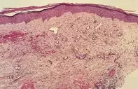
Acral angiodermatitis (pseudo Kaposi’s sarcoma) is the correct diagnosis in this case. The condition is characterised by slowly enlarging violaceous plaques which may closely mimic Kaposi’s sarcoma. Skin biopsy shows clusters of well differentiated vessels and fibrosis with haemorrhage and lymphocytic inflammation in the dermis.The process may complicate venous stasis, paralysis of the limbs, arteriovenous malformations or acquired arteriovenous fistulae, or it may appear on the stump of above-knee amputations. There is no effective treatment for acral angiodermatitis except minimising the limb oedema, and treating associated vascular disease.
Over a seven-year period, a 32-year-old man developed an increasing number of papules on the back of his calves which coalesced to form plaques (Figure 1). The plaques were asymptomatic and measured up to 11 cm in diameter. The process followed a bout of varicella pneumonia which was complicated by venous thromboses and persistent oedema of the legs. Skin biopsy showed clusters of well-differentiated vessels surrounded by dermal scarring and haemorrhage as well as haemosiderin pigment (Figure 2).

