What’s the diagnosis?
Infiltrated pretibial plaques


Chronic lymphoedema usually commences on the feet and is pitting. Biopsy shows no mucinosis.
Localised scleroderma may present as an infiltrated plaque but is usually unilateral and has an erythematous border. Skin biopsy shows extensive sclerosis of collagen and lymphocytic inflammation.
Lipodermatosclerosis is usually unilateral and is characterised by woody induration of the leg,. This is the result of chronic venous disease. Skin biopsy shows marked dermal and subcutaneous collagenous sclerosis with increased vessels as well as fibroblasts.
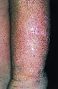
Pretibial myxoedema (thyroid dermopathy) is the correct diagnosis. It is usually seen in association with thyrotoxicosis and exophthalmos (Graves’ disease). In some patients the process may develop in conjunction with hypothyroidism or normal thyroid function. The fibroblasts in pretibial myxoedema have thyroid stimulating hormone receptors and are stimulated by circulating auto-antibodies to produce mucin. Topical high potency corticosteroid creams, intralesional corticosteroids or octreotide, surgery for polypoid nodules, and high dose intravenous immunoglobulin have been used for treatment. Complete resolution is uncommon.
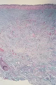
A 57-year-old woman developed infiltrated pretibial plaques on both legs. Over a period of three years the plaques had expanded onto her feet and the back of her calves (Figure 1). The plaques appeared waxy and were nonpitting. A skin biopsy showed marked dermal mucinosis in a greatly thickened skin (Figure 2). There was no associated inflammation.

