What’s the diagnosis?
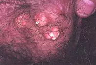
Multiple scrotal nodules with a chalky discharge


Multiple epithelial cysts may develop over the scrotum and range from flesh-colour to pale yellow to white smooth nodules. These cysts may discharge their keratinous contents into the surrounding tissue or onto the surface. Scrotal biopsy shows an epithelial cyst lined by stratified squamous epithelium and filled with loose, laminated keratin.
Sebaceous hyperplasia is often seen over the scrotum and is characterised by uniformly sized and distributed yellow papules. These may be highlighted when the scrotal skin is red and irritated. Scrotal biopsy shows clusters of mature sebaceous glands surrounding dilated follicular openings.
Molluscum contagiosum typically appears as multiple umbilicated papules with a firm pearly rim. Yellow to white grains may be extruded by curettage. Occasionally, molluscum contagiosum lesions may be quite large and inflamed. Skin biopsy shows lobular proliferation of the epidermis with numerous inclusion bodies representing aggregates of molluscum virus.
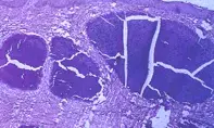
Idiopathic calcinosis of the scrotum is the correct diagnosis in this case. Scrotal biopsy revealed large amorphous deposits within the dermis that fractured on tissue sectioning (Figure 2). This condition appears in childhood or adulthood as either solitary or multiple asymptomatic nodules which may discharge a chalky material. The calcinosis is not associated with hypercalcaemia and represents a dystrophic calcification of scrotal epithelial cysts. The cyst lining may no longer be evident at the time of calcification. The cysts may rupture into the tissue inducing a foreign-body granulomatous reaction. Local curettage or surgical removal of symptomatic cysts is the treatment of choice.
A 45-year-old man presented with multiple asymptomatic nodules on his scrotum which discharged a chalky substance (Figure 1). The nodules had first appeared 10 years ago.

