What’s the diagnosis?
Persistent eroded lip in a young man

Allergic contact dermatitis may present as crusted and scaly lips. A localised plaque may be seen in individuals who suck metal- or rubber-tipped objects and in musicians. However, the dermatitis may extend beyond the vermilion border and involve both the upper and lower lips. Cosmetics, food substances or dentrifices may be further sources of allergens.
Irritant contact dermatitis (chapped lips) is common and is caused by a combination of environmental exposure (particularly chronic sun damage) and licking. The lips appear cracked and scaly and are often tender. Some forms may be complicated by haemorrhage and keratotic crusts and represent factitious self-induced cheilitis due to lip chewing. The use of vaseline-based moisturisers and sun-protection sticks is important in the management of this condition. Chapped lips are usually not infiltrated and do not pursue a relentless course.
Lupus erythematosus may rarely involve the lip and present as a localised hyperkeratotic and scaling plaque with a lacy, reticulate peripheral border. Skin biopsy shows an epidermis which is associated with basal cell layer damage and marked lymphocytic inflammation. Rarely, chronic plaques of lupus erythematosus may be complicated by the development of squamous cell carcinoma.
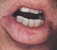
Squamous cell carcinoma is the correct diagnosis in this case, based on the clinical findings and on a biopsy, which was taken as the next step in management. The skin biopsy showed a hyperplastic epidermis with irregular lobular clusters of keratinocytes penetrating into the deep tissue (Figure 2). The keratinocytes showed a disorganised pattern of maturation and nuclear atypia, and there was premature keratinisation within the tumour lobules. The tumour arose in the background of an actinic cheilitis of the lower lip, which appeared mottled and pale with a loss of definition of the vermilion border. The diagnosis in this case was delayed because of the patient’s young age. The presence of a nonhealing infiltrating plaque which fails to respond to topical therapy should prompt skin biopsy, particularly in young individuals who have a history of chronic sun exposure in childhood or chronic occupational sun exposure. In this patient there was no evidence of lymph node spread and the tumour was successfully removed by surgery.
A 29-year-old man who worked as a builder developed an infiltrated and eroded 2.5 cm plaque on his lower lip (Figure 1). The plaque had gradually increased in size over a two-year period and had failed to heal with topical emollients, corticosteroids or antibacterial creams.

