What’s the diagnosis?
A persistent red nodule on the nose


Lymphocytoma cutis or lymphoma cutis may both present as red nodules. Skin biopsy shows sheets of lymphocytes with lymphoid follicles. Tests for clonal restriction of the lymphocytes may be required to confirm the presence of a lymphoma.
Granuloma faciale is a rare dermatological condition. It often presents on the nose as a reddish brown nodule or plaque. Skin biopsy shows a mixed inflammatory infiltrate with many eosinophils around the vessels in the upper dermis, and a lack of true granulomatous pathology, despite the name.
Lupus panniculitis represents a subcutaneous form of lupus erythematosus. This may present initially as a red nodule, which heals with atrophy due to fat loss. Skin biopsy shows deep hyaline fat necrosis with lymphoid inflammation but no granulomas.
Sarcoidosis (lupus pernio) is the correct diagnosis based on the biopsy result and subsequent review, which showed other similar cutaneous lesions, and a chest x-ray, which demonstrated bilateral hilar lymphadenopathy. Sarcoidosis is a multisystem granulomatous disease that in some patients may represent an exaggerated immunological reaction to mycobacteria antigens. Ophthalmic and pulmonary review are particularly important in assessing individual patients. Oral corticosteroids, antimalarials or methotrexate are used in treatment, depending on the severity and extent of the disease. For isolated cutaneous nodules, intralesional corticosteroids is an option.
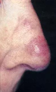
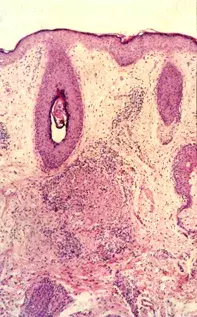
A 55-year-old woman gave an eight-month history of an asymptomatic red nodule on her nose (Figure 1). Skin biopsy showed a granuloma in the dermis together with a scant perivascular lymphocytic infiltrate (Figure 2). Polarisation of the tissue showed no foreign bodies and additional stains for organisms were negative.

