What’s the diagnosis?
Progressive dusky facial erythema


Rosacea may be associated with facial erythema and oedema. Rosacea is usually symmetrical and does not have a slowly progressive course. A history of recurrent flushing and the presence of pustules may be additional clues. Skin biopsy in rosacea shows telangiectasia, but not vascular proliferation.
Trauma may produce swelling and ecchymoses, but this is usually short lived and follows a course of progressive resolution, not progression.
Cellulitis may produce an asymmetrical, brawny swelling and erythema and has a very rapid course with fever. Cellulitis may be complicated by repeated episodes and persistent lymphoedema,
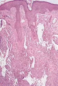
Angiosarcoma is the correct diagnosis in this case, based on the histopathology. It is a rare cutaneous malignancy seen primarily on the scalp and face in elderly individuals. Kaposi’s sarcoma is a variant of angiosarcoma, but it usually presents as multifocal disease and can usually be distinguished histologically. Angiosarcoma may also complicate postmastectomy lymphoedema (Stewart-Treve’s syndrome) or previous radiotherapy, particularly for breast cancer or childhood haemangioma. Early skin biopsy is the key to diagnosis. Limited tumours can be treated by surgery, but the majority will require radiotherapy or chemotherapy.
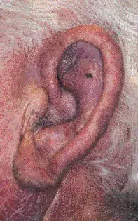
Over a 10-month period, a 77-year-old man developed a progressive dusky erythema and oedema on the left side of his face. This had commenced as a small plaque on the left cheek and had extended to the forehead, nose, left ear (Figure 1) and neck. The process had been asymptomatic. A skin biopsy showed irregular vascular channels, which extended into the deep dermis. The vessels were lined by enlarged endothelial cells which showed nuclear atypia and nuclear crowding, and there were detached endothelial cells in the vascular spaces (Figure 2).

