What’s the diagnosis?
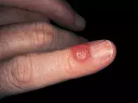
Translucent blister on the finger


Herpetic whitlow is painful, usually multilocular and lasts about three weeks. The blisters are initially serous but become pustular before resolving. Viral culture or cytological examination of the vesicle contents can confirm the diagnosis.
Insect bite may produce a solitary serous blister which is pruritic or painful and lasts a few days. Biopsy shows a pronounced inflammatory reaction with prominent eosinophils.
Orf or milker’s nodule may produce a solitary blister on the finger but this is usually purulent and is preceded by an inflamed nodule. A history of contact with farm animals potentially harbouring parapox virus may be obtained.
Myxoid (or mucous) cyst is the correct diagnosis in this case. These may be solitary or multiple. The mucous material is probably related to a leak of synovial fluid from the adjoining distal interphalangeal joint. The absence of an epithelial lining indicates that these are mucoceles rather than true cysts. Surgical removal, cryotherapy, intralesional corticosteroid injections (triamcinolone), aspiration and pressure dressing have all been used successfully in the treatment of myxoid cysts.
A 38-year-old man developed a painless 0.8 cm translucent blister proximal to the nail fold on his left index finger (Figure 1). Over a six-month period the blister intermittently discharged mucoid material but did not disappear. Biopsy showed pools of mucinous material in the dermis but no evident cystic lining (Figure 2).

