Dark follicular orifices
Skin lesions
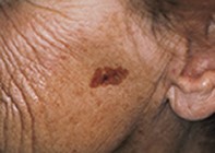
Case presentation
A 78-year-old woman presented with a lesion on her left cheek. It had been frozen nine years before the consultation and had subsequently recurred. The sharply demarcated lesion was dark tan and measured 2 cm x 1 cm (Figure 1). Dermatoscopy revealed multiple dark follicular orifices resembling pigment globules (Figure 2). These varied in size, and many were patulous. The surrounding background lesion had an orange–brown hue and a scalloped border. There was no pigment network visible. Skin biopsy revealed a hyperpigmented epidermis, but the number of melanocytes was normal and there was no atypia (Figure 3).
Single article purchases are temporarily unavailable due to site maintenance.
If you would like to purchase an article during this time, please email us at [email protected] with the article details and we'll assist you directly. We'll also let you know when online purchasing is available again.
Thank you for your patience and understanding.