Peer Reviewed
Perspectives on dermoscopy
Dermoscopic features of amelanotic and hypomelanotic melanoma
Recent articles on:
Melanoma
Melanoma
Recent articles on:
Skin cancer
Skin cancer
Abstract
Melanomas that lack significant pigment are a diagnostic challenge. Dermoscopy, although less accurate for diagnosing these lesions than for pigmented lesions, is still superior to clinical examination.
Key Points
Case presentations
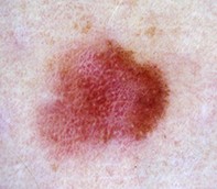
Case 1
A 34-year-old woman was referred by her GP for review of an asymptomatic pink lesion on her thigh. The main concern was that the lesion had grown slightly. She had no history of significant sun exposure. Clinical examination revealed a slightly elevated pink plaque (7 x 4 mm), which appeared relatively symmetrical (Figure 1a).
On dermoscopy, the lesion was a homogeneous pink colour with a central white area, most probably caused by the pressure of the non-polarised contact dermatoscope. Numerous dotted and linear irregular vessels were visible throughout the lesion (Figure 1b).
The lesion was excised with a 2 mm margin. Histopathological examination showed a 0.3 mm level II melanoma.
Purchase the PDF version of this article
Already a subscriber? Login here.