Bleeding varicose veins and a pustular rash
Test your diagnostic skills in our regular dermatology quiz. What is the cause of this pustular eruption?
Case presentation
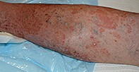
A 54-year-old man presents to the hospital emergency department concerned about bleeding from superficial varicose veins of his lower legs (Figure 1). Over the past week, he has developed a pustular rash. There is no itch or pain.
On examination, erythematous, moist, annular plaques are noted on the patient’s legs overlying the varicosities (Figure 2). Peripheral pulses are present. He also has pitting oedema to the knees.
The patient has a long history of recurrent cellulitis and venous stasis eczema and had been treated with long-term doxycycline and topical corticosteroid.
Differential diagnoses
Conditions to consider among the differential diagnoses include the following.
- Stasis dermatitis. This common condition (also known as varicose eczema) affects the lower limbs of patients with venous insufficiency. The skin is red to brown and may be firm to palpation, with overlying scale. The rash can be intensely pruritic. The case patient described above does have stasis dermatitis, but it is acutely exacerbated by a different skin disorder.
- Cellulitis and folliculitis. Cellulitis, which is usually caused by Streptococcus pyogenes, presents with a well-demarcated, erythematous, warm plaque typically located on the lower limb in adults. It is usually unilateral; bilateral cellulitis is generally secondary to a bilateral skin condition. Cellulitis is not pustular. Superficial pustules usually indicate concurrent folliculitis, an infection caused by Staphylococcus aureus.
- Pustular psoriasis. This acute skin condition may not have the same pathogenesis as psoriasis, with only 10% of affected patients having a background of psoriasis vulgaris. Pustular psoriasis has an acute onset and can be triggered by drugs (e.g. aspirin, indomethacin), infection or withdrawal of systemic corticosteroids. The rash is characterised by erythema, oedema and small sterile pustules. Patients with widespread pustular psoriasis may be unwell, with fever and rigors.
- Tinea corporis. This is the correct diagnosis. Tinea corporis is a dermatophyte infection that affects the keratinised surface of the skin on the trunk and extremities. In Australia, the most common cause is Trichophyton rubrum but other notable causes include Trichophyton tonsurans, Trichophyton mentagrophytes and Microsporum canis. Dogs, cats, cows and guinea pigs may be a source of tinea in people. However, tinea corporis in human adults is typically a result of secondary spread of T. rubrum from the feet (tinea pedis) or nails (tinea unguium). The presence of tinea on the feet and lower legs is a common portal of entry for streptococcal cellulitis.
Diagnosis
Microscopy and fungal culture are necessary for a diagnosis of tinea corporis because fungal infections frequently mimic many other skin conditions associated with scale, erythema and pustules. The plaque spreads centrifugally, so central sparing creates annular lesions with a raised scaly border. Pustules within the border are not common, so tinea presenting with peripheral pustules is often missed – as had occurred for this patient.1 However, tinea should always be considered in the differential diagnosis of a persistent pustular eruption, particularly when the result of bacterial culture is negative and there is no response to antibiotic therapy.
Management
Minor cutaneous dermatophyte infections are most commonly treated with topical terbinafine cream. Oral therapy is required for infections that are established, widespread or inflammatory, and also for lesions that have not responded to topical therapy.2 The most effective oral option is terbinafine (250 mg daily for two to three weeks), which has a fungicidal action. Other agents have fungistatic action: fluconazole and itraconazole are second-line agents, and griseofulvin is a third-line agent. Longer treatment periods are required for fungistatic agents (particularly for griseofulvin) and treatment needs to be continued until the rash has cleared and fungal scrapings are negative.
Patient education regarding foot hygiene is important to prevent further fungal infection. It is useful to instruct patients to apply topical antifungal treatment such as tolnaftate powder daily or terbinafine cream weekly to the feet and interdigital spaces. Although the rates of reinfection are high, a formal prophylactic regimen has not been developed.
Outcome
Bacterial swabs and fungal scrapings were taken from the pustular lesions for the case patient described above. A diagnosis of tinea corporis was confirmed by direct microscopy of a skin scraping from the border (prepared with potassium hydroxide) showing fungal hyphae; a culture from the scraping showed the definitive organism was T. rubrum. Swabs returned concurrent S. aureus.
The patient was treated with oral terbinafine 250 mg daily for three weeks. He was also managed with oral cephalexin and topical corticosteroids. The legs were elevated and wet dressings were applied, in addition to firm bandaging. As the inflammation decreased, the friability of his superficial varicose veins subsided. At the end of three weeks, fungal skin scrapings were negative and his skin had returned to normal. He was referred to a vascular surgeon, who confirmed venous insufficiency with ultrasound and placed the patient on a waiting list for bilateral saphenofemoral ligation and stripping of the varicose veins. He was also instructed to apply moisturiser to his legs and terbinafine cream to his feet and interdigital spaces weekly, and to continue this treatment indefinitely. MT
References

