A woman with multiple liver lesions
Liver diseases
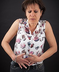
A 53-year-old woman with nausea and bloating of two months’ duration has multiple liver lesions detected on abdominal ultrasound. She is a new patient to your practice. How would you approach this situation?
Case scenario
Felicity is a 53-year-old teacher whose usual GP has recently retired. When she was last seen three weeks ago, she was experiencing nausea and bloating, which had persisted for two months. Physical examination was documented as normal and an upper abdominal ultrasound scan was requested. Her ultrasound demonstrates multiple solid liver lesions.
Commentary
Felicity has symptoms but liver lesions are often an incidental finding. In either case, when liver lesions are found it is essential to determine if these are benign or potentially more serious.
Cystic lesions and abscesses need to be differentiated from solid lesions. This is usually evident on ultrasound scanning, which is a common initial choice of imaging to investigate abdominal symptoms. Cysts require further investigation and treatment only if they are symptomatic, complex (e.g. calcified or septate) or multiple (more than five cysts may suggest the diagnosis of polycystic liver disease). In Felicity’s case, the lesions appear solid.
Hepatic haemangiomas are the most commonly detected benign solid liver lesion and are found three times more often in women than in men. Based on autopsy studies, the prevalence ranges from 0.4 to 20%.1 Although often solitary, multiple hepatic haemangiomas can occur. Other benign lesions include adenomas, focal nodular hyperplasia and regenerative nodules. Solid malignant lesions include hepatocellular carcinoma (HCC), metastases and cholangiocarcinoma.
An approach to investigating liver lesions is provided in the flowchart.
Important details from the history
Firstly, it should be established whether Felicity has had an ultrasound, CT scan or MRI of the liver in the past for comparison with the current findings. For example, if identical lesions were documented many years ago, they are unlikely to be malignant.
Eliciting a history of Felicity’s general health to assess the possibility of an underlying malignancy is essential. This should include, but not be limited to, asking about appetite, weight loss, bowel irregularity, headaches and cough. Gender and age of the patient are also relevant.
It is important to ask specifically about previous malignancies and underlying liver diseases, such as hepatitis B and C infection, nonalcoholic fatty liver disease, alcohol-related liver disease and haemochromatosis. Some malignancies, such as melanoma and breast cancer, may recur many years after their initial diagnosis.
Smoking and alcohol history should be obtained. Use of the oral contraceptive pill is relevant because long-term use has been associated with hepatic adenomas. A thorough physical examination should also be undertaken, including a search for breast lumps and lymphadenopathy.
Which blood tests are useful?
Liver biochemistry may provide clues. An isolated elevated serum alkaline phosphatase level may occur with liver as well as bone metastases. It may also be found in unrelated conditions, such as Paget’s disease. An elevated alkaline phosphatase level together with an elevated level of gamma glutamyl transferase (GGT) can indicate cholestasis; however, GGT is nonspecific and may be secondary to hepatic infiltration with fat, medications, drugs or inflammation. Causes of cholestasis include biliary obstruction and hepatic infiltration as well as the two chronic cholestatic liver diseases: primary biliary cholangitis and primary sclerosing cholangitis.
A markedly elevated alpha-fetoprotein (AFP) level in the presence of cirrhosis is highly suggestive of hepatocellular carcinoma (HCC), but only about 60% of HCCs result in a raised AFP level. Other tumour markers are of limited value. Cancer antigen 19-9 (CA 19-9), carcinoembryonic antigen (CEA) and carcinoma antigen 15-3 (CA 15-3) levels may be elevated, most often in upper gastrointestinal, colorectal and breast cancer, respectively. However, these are nonspecific markers and initial testing for these is not encouraged for diagnosis.
Further imaging
Further imaging is often necessary. Many liver lesions have a characteristic appearance; therefore, choice of imaging modality should be based on the likely diagnosis. In a noncirrhotic liver, haemangiomas and adenomas generally have a typical appearance on multiphase CT scanning or contrast- enhanced MRI. These scans may also reveal a central scar, which is characteristic of focal nodular hyperplasia.
When cirrhosis is present, a liver mass or nodule should be assumed initially to be an HCC.2 Multiphase CT and MRI scans are often diagnostic (obviating the need for biopsy) when the lesion is greater than 1 cm in diameter. Metastases are generally less vascular than HCC. Positive emission tomography using fluorodeoxyglucose as the tracer (FDG-PET) is sensitive for detecting many malignancies but is not always available or reimbursable; its use in diagnosis of liver metastases is limited.
When there is doubt, MRI using a hepatobiliary contrast agent such as primovist is useful for distinguishing between adenomas and focal nodular hyperplasia. The use of labelled red blood cell scans for diagnosing haemangiomas has largely been superseded, although this test may have a role in diagnosing atypical lesions.
When the diagnosis is unclear
Although many liver lesions have characteristic radiological appearances, not uncommonly a definitive diagnosis is unable to be reached, particularly if the lesions are less than 1 cm in diameter. When the diagnosis is unclear, referral to a gastroenterologist or upper gastrointestinal surgeon should be considered, particularly if there is a strong suspicion of malignancy. However, observation and monitoring with serial ultrasound may be all that is required.
In some cases, a specialist may decide to perform a targeted biopsy, providing the lesion is in a suitable position. Rarely, excision biopsy will be considered, particularly when the lesion is suspicious for a malignancy and not in an easy position to biopsy percutaneously. Potentially curable lesions diagnosed by imaging characteristics, such as HCC, can be resected without biopsy.2
Conclusion
In patients such as Felicity with multiple solid hepatic lesions, the first consideration is to exclude malignancy. In many cases, the diagnosis can be reached noninvasively.3 A starting point is viewing previous imaging, taking a focused history, performing a targeted examination and reviewing liver biochemistry test results. Choice of additional imaging modality should be guided by the provisional diagnosis. MT
References
- Bajenaru N, Balaban V, Savulescu F, Campeanu I, Patrascu T. Hepatic hemangioma – review. J Med Life 2015; 8(Spec Issue): 4-11.
- Marrero JA, Ahn J, Reddy KR; Practice Parameters Committee of the American College of Gastroenterology (ACG). ACG clinical guidelines: the diagnosis and management of focal liver lesions.
- Am J Gastroenterol 2014; 109: 1328-1347. Available at http://gi.org/wp-content/uploads/2014/08/ACG_Guideline_Focal_Liver_Lesions_September_2014.pdf (accessed May 2017).
- Schwartz JM, Kruskal J. Solid liver lesions: differential diagnosis and evaluation UpToDate April 2017. Available at http://www.uptodate.com/contents/solid-liver-lesions-differential-diagnosis-and-evaluation (accessed May 2017).
COMPETING INTERESTS: None.