An atypical mole
Skin lesions
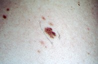
Case presentation
A 34-year-old man had at least 10 atypical benign moles removed over an 18-year period. At the time of consultation he had a 7 mm diameter, irregularly shaped mole of unknown duration on his upper right back (Figure 1). Dermoscopy showed a patchy pigment network mixed with scattered pigment globules and dots. The network was accentuated in two regions, but the pigment was not coarse and there were no peripheral streams of pigment or blunt pseudopods (Figure 2). Excision biopsy revealed an epidermis with a prominent and irregular rete ridge system. There were nests of melanocytes at the tips of some of the rete ridges, while other rete tips were spared. Nests of uniform melanocytes were also evident in the upper dermis (Figure 3). There was no melanocytic atypic.