Peer Reviewed
Perspectives on dermoscopy
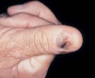
Progressively pigmented thumb nail
Recent articles on:
Melanoma
Melanoma
Abstract
Another case in our series, to help you hone your skills in dermoscopy.
Key Points
Case presentation
Over an eight-year period, a 60-year-old man noted progressive dark discolouration of his thumb nail with disintegration of the distal nail plate (Figure 1). Dermoscopy revealed broad bands of dark pigment within the nail bed and finer radiating pigmented streaks extending into the surrounding skin (Figure 2). There were scattered pigment dots and a patchy grey–white milky veil. Excision revealed a confluent proliferation of atypical melanocytes that were intraepidermal and associated with superficial lymphocytic inflammation and fibrosis (Figure 3).
Purchase the PDF version of this article
Already a subscriber? Login here.