Peer Reviewed
Perspectives on dermoscopy
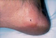
A pigmented lesion on the sole
Recent articles on:
Skin lesions
Skin lesions
Abstract
Dermoscopy is useful in evaluating small pigmented lesions on the sole, but some lesions may show areas of asymmetric homogenous pigment, prompting biopsy.
Key Points
Case presentation
A 39-year-old woman noted an asymmetrical dark mole over the medial aspect of her right posterior sole (Figure 1). The mole had appeared over the previous 12 months and measured 3 mm in diameter. Dermoscopy showed a lattice-like pigment network with small brown globules. In the upper pole of the lesion the network was obscured by larger blue–black globules (Figure 2). Excision biopsy revealed a well developed epidermal rete ridge system with nests of melanocytes at their tips and also within the superficial dermis. The junctional and superficial dermal nests were deeply pigmented, but there was no atypia (Figure 3).