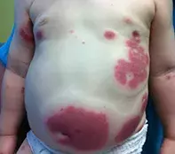
What’s the diagnosis?
A child with a palpable and purpuric eruption

Case presentation
An 8-month-old boy presents with an oedematous and purpuric eruption affecting the trunk (Figure). He has had a mild fever for the past 24 hours, but at presentation he appears to be happy and active.
Differential diagnosis
Conditions to consider in the differential diagnosis for a child of this age include the following.
- Henoch–Schönlein purpura. This is the most common type of small vessel vasculitis in children and is characterised by a palpable, erythematous purpura that typically affects the lower limbs and buttocks. It is sometimes accompanied by oedema and often by arthralgia, gastritis and nephritis. Although largely a clinical diagnosis, Henoch–Schönlein purpura can be confirmed by skin biopsy, which will show IgA immune complex deposition in the walls of arterioles. Active treatment is not always required. A small proportion of children (<5%) are at increased risk of developing renal disease later in life and so a urinalysis should be performed for all patients who are suspected of having this condition.
- Common urticaria. This very common acute vascular reaction of the skin is driven by a histamine release that causes arteriolar vasodilatation and increased capillary permeability. It is most often associated with a viral illness, but medications or environmental triggers may be responsible in young children. Lesions are typically raised erythematous wheals that are variable in shape, size and location. Classic features include lesion migration, no fixed distribution and intense pruritus (although in children it can be surprisingly asymptomatic). Although there may be a slightly bruised or purple appearance in a young child, frank purpura is never present in common urticaria. The condition is usually self-limiting. Antihistamines are contraindicated in patients under 12 months of age but useful to suppress the itchy rash in older children.
- Erythema multiforme. This is an acute immune-mediated cutaneous reaction secondary to a specific antigen (most commonly to herpes simplex virus or Mycoplasma pneumoniae; more rarely to medications). Repeated attacks can occur when it is triggered by herpes simplex virus. The lesions have a distinctive targetoid appearance – a focus of redness surrounded by a grey discolouration and a red outer ring – but oedema is not a feature. Sometimes there is a vesiculobullous component with erosions. Erythema multiforme commonly involves the extremities, such as the palms and soles, and the mucosal surfaces of the genitalia, oral cavity and conjunctiva; the distribution of the lesions shown in the Figure would be atypical for erythema multiforme. Usually the illness is benign but occasionally it is severe, with extreme mucosal involvement and ocular involvement that may lead to visual loss.
- Viral exanthem. Viral illnesses can manifest as a palpable purpuric skin eruption, with or without a blistering component. They are accompanied by other systemic clinical signs, particularly fever and malaise, which are often mild. Viruses that are commonly responsible include human herpes virus types 6 and 7 (roseola infantum), parvovirus B19 (erythema infectiosum), Epstein Barr virus, cytomegalovirus, and hepatitis B and C viruses.
- Acute haemorrhagic oedema. This small-vessel leukocytoclastic vasculitis is the correct diagnosis. It is characterised by mild pyrexia and large tender wheals that are oedematous and purpuric. The cause is unknown, but it is thought to be triggered by infection or medications. The condition progresses rapidly over 24 to 48 hours, and the oedema occurs within the haemorrhagic ecchymotic plaques and can extend beyond these. It typically occurs on the face and limbs but, as seen in the case above, it can also affect the truncal region. Acute haemorrhagic oedema is most common in children aged between 4 months and 2 years, and affected individuals tend to be otherwise systemically well at presentation.
Investigations
Acute haemorrhagic oedema of infancy is a clinical diagnosis. However, if the diagnosis is not entirely clear then basic tests can be conducted to exclude other illness and systemic organ involvement (full blood count, C-reactive protein [CRP], erythrocyte sedimentation rate [ESR], electrolytes, with kidney function and a urinalysis). Blood tests may show an increased ESR and a leucocytosis, and there may be evidence of proteinuria and haematuria. A biopsy is not necessary but if taken will show evidence of inflammatory blood vessels with white cell destruction and an absence of IgA immune complexes.
Joint, renal and gastrointestinal involvement has been described for patients with acute haemorrhagic oedema but is very uncommon (and reported cases may in fact have been Henoch–Schönlein purpura). The two conditions can be difficult to distinguish – when there is a suspicion of Henoch–Schönlein purpura a urinalysis should be performed.
Management
Acute haemorrhagic oedema resolves spontaneously within two weeks and no particular treatment is required. Some lesions may recur intermittently, but there are no significant long term sequelae.

